
Sphincter
| Sphincter | |
|---|---|
| Identifiers | |
| TA98 | A04.0.00.028 |
| TA2 | 1992 |
| FMA | 75004 |
| Anatomical terms of muscle | |

A sphincter is a circular muscle that normally maintains constriction of a natural body passage or orifice and relaxes as required by normal physiological functioning. Sphincters are found in many animals. There are over 60 types in the human body, some microscopically small, in particular the millions of precapillary sphincters.[1] Sphincters relax at death, often releasing fluids and feces.[2]
Functioning
Each sphincter is associated with the lumen (opening) it surrounds. As long as the sphincter muscle is contracted, its length is shortened and the lumen is constricted (closed). Relaxation of the muscle causes it to lengthen, opening the lumen and allowing the passage of liquids, solids, or gases.
This is evident, for example, in the blowholes of numerous marine mammals.
Many sphincters are used every day in the normal course of digestion. For example, the lower esophageal sphincter (or cardiac sphincter), which resides at the top of the stomach, is closed most of the time, keeping acids and other stomach contents from pushing up and into the oesophagus, but opens to let swallowed food pass into the stomach.
Classifications
Sphincters can be further classified into functional and anatomical sphincters:
- Anatomical sphincters have a localised and often circular muscle thickening to facilitate their action as a sphincter.
- Functional sphincters do not have this localised muscle thickening and achieve their sphincteric action through muscle contraction around (extrinsic) or within (intrinsic) the structure.
Sphincters can also be voluntarily or involuntarily controlled:
- Voluntary sphincters are supplied by somatic nerves.
- Involuntary sphincters are stimulated by autonomic nerves.
Examples
- The pupillary sphincter, belonging to the iris in the eye.
- The orbicularis oculi muscle, a sphincter-like muscle around the eye.
- The upper oesophageal sphincter
- หูรูดหลอดอาหารส่วนล่างหรือหูรูดหัวใจ อยู่ที่ส่วนบน ( คาร์เดีย ) ของกระเพาะอาหารหูรูดนี้ป้องกันไม่ ให้ กรด ในกระเพาะ อาหารไหลย้อนขึ้นไปในหลอดอาหาร
- หูรูดไพลอริกซึ่งอยู่บริเวณส่วนล่างของกระเพาะอาหาร
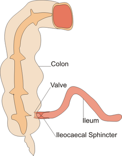
- หูรูดอิเลโอซีคัล (ileocecal sphincter)บริเวณรอยต่อระหว่างลำไส้เล็ก (ileum) และลำไส้ใหญ่ทำหน้าที่จำกัดการไหลย้อนกลับของสารคัดหลั่งจากลำไส้ใหญ่กลับเข้าไปในลำไส้เล็ก
- หูรูดของออดดีหรือหูรูดของกลิสสัน ทำหน้าที่ควบคุมการหลั่งสารจากตับตับอ่อนและถุงน้ำดีเข้าสู่ลำไส้เล็กส่วนต้น
- กล้ามเนื้อหูรูดท่อปัสสาวะทำหน้าที่ควบคุมการขับปัสสาวะ ออก จากร่างกาย
- บริเวณทวารหนักมีกล้ามเนื้อหูรูดทวารหนักสองมัดที่ควบคุมการขับถ่ายอุจจาระ ออก จากร่างกาย ได้แก่กล้ามเนื้อหูรูดทวารหนักส่วนในและกล้ามเนื้อหูรูดทวารหนักส่วนนอก กล้ามเนื้อหูรูดส่วนในทำงาน โดยไม่รู้ตัวส่วนกล้ามเนื้อหูรูดส่วนนอกทำงานโดยรู้ตัว
- หูรูดก่อนเส้นเลือดฝอยขนาดเล็กทำหน้าที่ควบคุม การไหลเวียน ของเลือด เข้าสู่ เส้นเลือดฝอยแต่ละเส้นเพื่อตอบสนองต่อกิจกรรมการเผาผลาญ ในบริเวณนั้น [ 1 ]
- กล้ามเนื้อ ออร์บิคูลาริส โอริส (ไม่ใช่กล้ามเนื้อหูรูดแท้จริง) เป็นกลุ่มกล้ามเนื้อในริมฝีปากที่ล้อมรอบปาก
ความสำคัญทางคลินิก
มีการผ่าตัดที่เรียกว่าsphincterotomyซึ่งเป็นการตัดกล้ามเนื้อหูรูดเพื่อบรรเทาอาการต่างๆ การผ่าตัดเหล่านี้บางส่วนได้แก่lateral internal sphincterotomyและanal sphincterotomyเพื่อรักษาแผลปริที่ทวารหนัก [ 3 ]และbiliary endoscopic sphincterotomyเพื่อรักษาภาวะทางเดินอาหารบางอย่าง รวมถึงการเอาหินน้ำดี ออก