
กล้ามเนื้อ Vastus
กล้ามเนื้อวาสตัส (Vastus muscles)เป็นหนึ่งในสี่กล้ามเนื้อที่ประกอบกันเป็นกล้ามเนื้อควอดริเซปส์ เฟโมริส (Quadriceps femoris muscle ) บริเวณ ต้น ขากล้ามเนื้อทั้งสามมัดได้แก่ วาสตัส อินเตอร์มีเดีย ส (Vastus intermedius ), วาสตัส ลาเทอราลิส (Vastus lateralis ) และ วาสตั ส มีเดียลิส (Vastus medialis)ซึ่งตั้งอยู่ตรงกลาง ด้านนอก และด้านในของต้นขาตามลำดับ ส่วนกล้ามเนื้อมัดที่สี่คือเรคตัส เฟโมริส (Rectus femoris)ซึ่ง เป็น กล้ามเนื้อขนาดใหญ่ที่คลุมอยู่ด้านหน้าและด้านข้างของกระดูกต้นขา
วาสตัส อินเตอร์มีเดียส
กล้ามเนื้อ วาสตัส อินเตอร์มีเดียส (Vastus intermedius) เกิดจากพื้นผิวด้านหน้าและด้านข้างของตัวกระดูกต้นขาในส่วนบนสองในสาม โดยอยู่ใต้กล้ามเนื้อเรคตัส เฟโมริส (Rectus femoris) และจากส่วนล่างของเยื่อกั้นกล้ามเนื้อด้านข้าง (Lateral intermuscular septum ) เส้นใยของมันสิ้นสุดลงที่พังผืดชั้นตื้น (Superficial aponeurosis ) ซึ่งก่อตัวเป็นส่วนลึกของเอ็นควอดริเซปส์ (Quadriceps tendon )
กล้ามเนื้อวาสตัสมีเดียลิสและวาสตัสอินเตอร์มีเดียสดูเหมือนจะเชื่อมต่อกันอย่างแยกไม่ออก แต่เมื่อกล้ามเนื้อเรคตัสเฟโมริสถูกเลื่อนออกไปจะสังเกตเห็นช่องว่างแคบๆ ที่ทอดยาวขึ้นมาจาก ขอบ ด้านในของกระดูกสะบ้าระหว่างกล้ามเนื้อทั้งสอง และการแยกนี้อาจต่อเนื่องไปจนถึงส่วนล่างของเส้นอินเตอร์โทรแคนเทอริกซึ่งในบริเวณนั้น กล้ามเนื้อทั้งสองมักจะเชื่อมต่อกัน
วาสตัส ลาเทอราลิส
กล้ามเนื้อวาสตัส ลาเทอราลิส (Vastus lateralis) เป็นกล้ามเนื้อที่ใหญ่ที่สุดและแข็งแรงที่สุดในบรรดากล้ามเนื้อวาสติทั้งสามมัด มันเริ่มต้นจากหลายส่วนของกระดูกต้นขา รวมถึงส่วนบนของเส้นอินเตอร์โทรแคนเทอริก (Intertrochanteric line) ขอบด้านหน้าส่วนล่าง ของกระดูก โทรแคนเตอร์ใหญ่ (Greater Trochanter)ไปจนถึงขอบด้านนอกของปุ่มกระดูกกลูเตียล (Gluteal tuberosity ) และครึ่งบนของขอบด้านนอกของเส้นลิเนีย แอสเปรา (Linea aspera ) ส่วนต่างๆ เหล่านี้รวมตัวกันเป็นเอ็นอะพอนิวโรซิส (Aponeurosis) ซึ่งเป็นเอ็นแบนกว้างที่คลุมส่วนบนสามในสี่ของกล้ามเนื้อ จากพื้นผิวด้านในของเอ็นอะพอนิวโรซิส มีเส้นใยกล้ามเนื้อจำนวนมากเกิดขึ้น นอกจากนี้ยังมีเส้นใยเพิ่มเติมบางส่วนที่เกิดขึ้นจากเอ็นของกล้าม เนื้อ กลูเตียส แม็กซิมัส (Gluteus maximus ) และจากเยื่อกั้นระหว่างกล้ามเนื้อวาสตัส ลาเทอราลิส กับส่วนหัวสั้นของ กล้ามเนื้อ ไบเซปส์ เฟโมริส (Biceps femoris )
เส้นใยเหล่านี้รวมตัวกันเป็นก้อนเนื้อขนาดใหญ่ ยึดติดกับพังผืดที่แข็งแรงอีกอันหนึ่ง ซึ่งอยู่บนพื้นผิวด้านลึกของส่วนล่างของกล้ามเนื้อ พังผืดด้านล่างนี้จะหดตัวและหนาขึ้นกลายเป็นเอ็นแบนที่ยึดติดกับขอบด้านนอกของกระดูกสะบ้าและต่อมาเชื่อมต่อกับเอ็นของกล้ามเนื้อต้นขาด้านหน้า (quadriceps femoris tendon ) ทำให้ แคปซูลของข้อเข่าขยายออก
วาสตัส มีเดียลิส
The vastus medialis arises medially along the entire length of the femur, and attaches with the other muscles of the quadriceps in the quadriceps tendon.[1] The vastus medialis muscle originates from a continuous line of attachment on the femur, which begins on the front and middle side (anteromedially) on the intertrochanteric line of the femur. It continues down and back (posteroinferiorly) along the pectineal line and then descends along the inner lip of the linea aspera and onto the medial supracondylar line of the femur. The fibers converge onto the medial part of the quadriceps tendon and the medial border of the patella.[1] The obliquus genus muscle is the most distal segment of the vastus medialis muscle. Its specific training plays an important role in maintaining patella position and limiting injuries to the knee. With no clear delineation, it is simply the most distal group of fibers of the vastus medialis.
Additional images
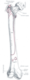 Right femur. Anterior surface.
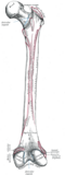 Right femur. Posterior surface.
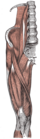 Muscles of the iliac and anterior femoral regions.
 Cross-section through the middle of the thigh.
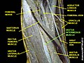 Vastus intermedius muscle
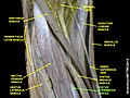 Vastus intermedius muscle
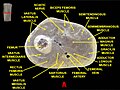 Muscles of thigh. Cross section.
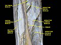 Muscles of Thigh. Anterior views.