
Sialography
| Sialography | |
|---|---|
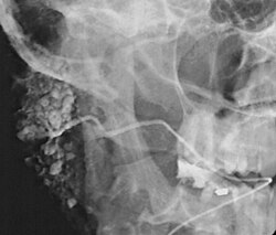 Sialogram in a patient suspected of Sjögren's syndrome | |
| MeSH | D012796 |
| MedlinePlus | 003812 |
Sialography (also termed radiosialography) is the radiographic examination of the salivary glands. It usually involves the injection of a small amount of contrast medium into the salivary duct of a single gland, followed by routine X-ray projections.[1]
The resulting image is called a sialogram.
Sialography has largely been replaced by sialoendoscopy[2] and cross-sectional imaging, such as CT, MRI and ultrasonography.[3]
Uses
This procedure is indicated when there is recurrent swelling and pain on the face but ultrasound has not revealed any problems. If Sjögren syndrome (also known as Sicca syndrome, an autoimmune disease that affects the lacrimal and salivary glands, causing reduced tears and saliva production) is suspected, this procedure is useful. Besides, when interventional procedure is planned such as stone removal from salivary ducts or dilatation of the strictures in the salivary gland, this procedure is also indicated.[4] However, for those who are pregnant, with allergy to iodinated contrast, and ongoing infection or inflammation of the face, the procedure is contraindicated.[4]
Procedure
Contrast agents are classified into two groups: fat-soluble contrast agents and water-soluble contrast agents. Water-soluble contrast agents can fill the finer elements of the ductal system. Fat-soluble contrast agents are viscous and can cause allergic reactions. These can also cause discomfort to the patients. Fat-soluble contrast agents do not fill finer elements of the duct.
A baseline radiograph (scout film) of the required salivary gland would be taken, the duct is dilated using graded lacrimal probes, a cannula then is inserted in this salivary gland duct's opening in the mouth, then a radio-opaque fluid (contrast medium) is injected in the duct through a small tube.
A series of radiographs would then be taken to determine the flow of the fluid, identify any obstructions and its location, the rate of fluid excretion from the gland.
Usually the radiographs taken are lateral oblique views of the face[5] as orthopantomograms are not useful for the purpose of locating the area due to superimpositions and the way they are taken to put the teeth in the main field.
Interpretation
การศึกษานี้ตีความโดยการประเมินสัณฐานวิทยาของท่อต่อมน้ำลายเพื่อตรวจหาการอุดตันและการอักเสบเรื้อรัง ภาวะท่อต่อมน้ำ ลายอักเสบ ( Sialodochitis ) เป็นคำที่ใช้อธิบายการขยายตัวของท่อที่เกิดจากกระบวนการอักเสบหรือติดเชื้อซ้ำๆ นอกจากนี้ยังพบภาวะตีบแคบของท่อต่อมน้ำลายที่ไม่สม่ำเสมอ ซึ่งทำให้เกิดลักษณะที่เรียกว่า "รูปแบบไส้กรอก" บนภาพเอกซเรย์ต่อมน้ำลาย อาจพบสัญญาณของฝีและโรคภูมิต้านตนเอง เช่น กลุ่มอาการ Sjögren ภาวะ ต่อมน้ำลาย อักเสบ (Sialadenitis)คือการอักเสบของต่อมน้ำลาย ซึ่งอาจทำให้เกิดการฝ่อของอะซินาร์และทำให้เกิดลักษณะที่เรียกว่า "การตัดแต่งกิ่งของต้นไม้" บนภาพเอกซเรย์ต่อมน้ำลาย โดยมีกิ่งก้านที่มองเห็นได้จากระบบท่อน้อยลง รอยโรคที่กินพื้นที่ภายในหรือติดกับต่อมน้ำลายอาจทำให้โครงสร้างทางกายวิภาคปกติของต่อมเปลี่ยนไป สิ่งนี้อาจทำให้เกิดลักษณะที่เรียกว่า "ลูกบอลในมือ" บนภาพรังสีต่อมน้ำลาย โดยที่ท่อจะโค้งรอบก้อนเนื้อ[ 6 ]
ผลข้างเคียง
เช่นเดียวกับการถ่ายภาพทางการแพทย์ใดๆ ที่ใช้รังสีไอออนไนซ์จะมีทั้งความเสียหายโดยตรงจากรังสีไอออนไนซ์และความเสียหายทางอ้อมจากอนุมูลอิสระที่เกิดขึ้นระหว่างการแตกตัวเป็นไอออนของโมเลกุลน้ำภายในเซลล์ ความเสี่ยงที่จะทำให้เกิดเนื้องอกร้ายนั้นน้อยมาก และจะถูกพิจารณาเทียบกับประโยชน์ของการตรวจวินิจฉัยเป็นรายกรณีไป
ภาวะแทรกซ้อนที่อาจเกิดขึ้นได้ ได้แก่:
- เจ็บขณะฉีดยา
- การติดเชื้อหลังการทำหัตถการ
- ท่อแตก
- การรั่วไหลของสารทึบแสง
- อาการแพ้สารทึบรังสีไอโอดีน (ซึ่งอาจแสดงอาการลมพิษหายใจลำบากและความดันโลหิตต่ำ )