อ่าน 35 นาที
Artificial cell
CS1 แหล่งที่มาภาษาเยอรมัน (de)/ชีววิทยาของเซลล์/ชีววิทยาสังเคราะห์
An artificial cell, synthetic cell or minimal cell is an engineered particle that mimics one or many functions of a biological cell.
Artificial cell
An artificial cell, synthetic cell or minimal cell is an engineered particle that mimics one or many functions of a biological cell. Often, artificial cells are biological or polymeric membranes which enclose biologically active materials.[1] As such, liposomes, polymersomes, nanoparticles, microcapsules and a number of other particles can qualify as artificial cells.
The terms "artificial cell" and "synthetic cell" are used in a variety of different fields and can have different meanings. Some stricter definitions are based on the assumption that the term "cell" directly relates to biological cells and that these structures therefore have to be alive (or part of a living organism) and, further, that the term "artificial" implies that these structures are artificially built from the bottom-up, i.e. from basic components. As such, in the area of synthetic biology, an artificial cell can be understood as a completely synthetically made cell that can capture energy, maintain ion gradients, contain macromolecules as well as store information and have the ability to replicate.[2] This kind of artificial cell has not yet been made.
Alternatively, the term "artificial" does not imply that the entire structure is man-made, but instead, it can refer to the idea that certain functions or structures of biological cells can be modified, simplified, replaced or supplemented with a synthetic entity.
In other fields, the term "artificial cell" can refer to any compartment that somewhat resembles a biological cell in size or structure, but is synthetically made, or even fully made from non-biological components. The term "artificial cell" is also used for structures with direct applications such as compartments for drug delivery. Micro-encapsulation allows for metabolism within the membrane, exchange of small molecules and prevention of passage of large substances across it.[3][4] The main advantages of encapsulation include improved mimicry in the body, increased solubility of the cargo and decreased immune responses. Artificial cells have been clinically successful in hemoperfusion.[5]
| Part of a series of articles on |
| Synthetic biology |
|---|
| Synthetic biological circuits |
| Genome editing |
| Artificial cells |
| Xenobiology |
| Other topics |
Bottom-up engineering of living artificial cells

German pathologist Rudolf Virchow proposed that not only does life arise from cells, but every cell comes from another cell; "Omnis cellula e cellula".[6] Until now, most attempts to create an artificial cell have engineered modules that can mimic certain functions of living cells. Advances in cell-free transcription and translation reactions allow the expression of many genes as well as interdependent genetic and metabolic networks,[7] but these efforts are still far from producing a fully operational cell.
A bottom-up approach to build an artificial cell would involve creating a protocellde novo, entirely from non-living materials. As the term "cell" implies, one prerequisite is the generation of some sort of compartment that defines an individual, cellular unit. Phospholipid membranes are an obvious choice as compartmentalizing boundaries,[8] as they act as selective barriers in all living biological cells. Scientists can encapsulate biomolecules in cell-sized phospholipid vesicles and by doing so, observe these molecules to act similarly as in biological cells and thereby recreate certain cell functions.[9] In a similar way, functional biological building blocks can be encapsulated in these lipid compartments to achieve the synthesis of (however rudimentary) artificial cells.
Researchers have proposed creating a phospholipid bilayer vesicle with DNA capable of self-reproducing using synthetic genetic information. The three primary elements of such artificial cells are the formation of a lipid membrane, DNA and RNA replication through a template process and the harvesting of chemical energy for active transport across the membrane.[10][11] The main hurdles foreseen and encountered with this proposed protocell are the creation of a minimal synthetic DNA that holds all sufficient information for life, and the reproduction of non-genetic components that are integral in cell development such as molecular self-organization.[12] However, it is hoped that this kind of bottom-up approach would provide insight into the fundamental questions of organizations at the cellular level and the origins of biological life. So far, no completely artificial cell capable of self-reproduction has been synthesized using the molecules of life, and this objective is still in the distant future although various groups are currently working towards this goal.[13]
Another method proposed to create a protocell more closely resembles the conditions believed to have been present during evolution known as the primordial soup. Various RNA polymers could be encapsulated in vesicles and in such small boundary conditions, chemical reactions would be tested for.[14]
Ethics and controversy
Protocell research has created controversy and opposing opinions, including critics of the vague definition of "artificial life".[15] The creation of a basic unit of life is the most pressing ethical concern.[16] Synthetic organisms could escape and cause damage to human health and ecosystems, or the technology could be used to make a biological weapon.[17] Cells with certain non-standard biochemistries, such as mirror life, could also have a competitive advantage over natural organisms.[18] Although according to Ting Zhu, "the creation of a mirror-image organism lies well beyond the reach of present-day science".[19]
International research community
In the mid-2010s the research community started recognising the need to unify the field of synthetic cell research, acknowledging that the task of constructing an entire living organism from non-living components was beyond the resources of a single country.[20]
ในปี 2017 โครงการความร่วมมือวิจัยขนาดใหญ่ระดับนานาชาติBuild-a-Cell ที่ได้รับทุนสนับสนุนจาก NSF สำหรับการสร้างเซลล์สิ่งมีชีวิตสังเคราะห์ได้เริ่มต้นขึ้น [ 21 ] Build-a-Cell ได้จัดกิจกรรมการประชุมเชิงปฏิบัติการแบบสหวิทยาการจำนวน 9 ครั้ง ซึ่งเปิดให้ผู้สนใจทุกคนเข้าร่วม เพื่อหารือและชี้นำอนาคตของชุมชนเซลล์สังเคราะห์ Build-a-Cell ได้รับการสานต่อโดยองค์กรเซลล์สังเคราะห์ระดับชาติในหลายประเทศ องค์กรระดับชาติเหล่านั้นได้แก่ FabriCell [ 22 ] MaxSynBio [ 23 ]และ BaSyC [ 24 ]ความพยายามด้านเซลล์สังเคราะห์ของยุโรปได้รวมกันในปี 2019 ในชื่อโครงการ SynCellEU [ 25 ]
วิธีการสร้างเซลล์สิ่งมีชีวิตขนาดเล็กโดยใช้แนวทางจากบนลงล่าง
สมาชิกจากสถาบัน J. Craig Venterได้ใช้ วิธีการคำนวณ แบบบนลงล่างเพื่อกำจัดยีนในสิ่งมีชีวิตให้เหลือชุดยีนขั้นต่ำ[ 26 ]ในปี 2010 ทีมงานประสบความสำเร็จในการสร้างสายพันธุ์ที่จำลองตัวเองได้ (ชื่อMycoplasma laboratorium ) ของMycoplasma mycoidesโดยใช้ DNA ที่สร้างขึ้นสังเคราะห์ซึ่งถือว่าเป็นข้อกำหนดขั้นต่ำสำหรับการดำรงชีวิต ซึ่งถูกแทรกเข้าไปในแบคทีเรียที่ไม่มีจีโนม[ 26 ]ผู้สนับสนุนแนะนำว่ากระบวนการสังเคราะห์ทางชีวภาพแบบบนลงล่างจะช่วยให้สามารถแทรกยีนใหม่ที่จะทำหน้าที่ที่เป็นประโยชน์ เช่น การสร้างไฮโดรเจนสำหรับเชื้อเพลิงหรือการดักจับคาร์บอนไดออกไซด์ส่วนเกินในชั้นบรรยากาศ[ 16 ]เครือข่ายการควบคุม การเผาผลาญ และการส่งสัญญาณจำนวนมากยังไม่ได้รับการระบุลักษณะอย่างสมบูรณ์ วิธีการแบบ บนลงล่าง เหล่านี้ มีข้อจำกัดในการทำความเข้าใจการควบคุมโมเลกุลพื้นฐาน เนื่องจากสิ่งมีชีวิตเจ้าบ้านมีองค์ประกอบโมเลกุลที่ซับซ้อนและไม่ได้กำหนดไว้อย่างสมบูรณ์[ 27 ]ในปี 2019 ได้มีการเผยแพร่แบบจำลองการคำนวณที่สมบูรณ์ของเส้นทางทั้งหมดในเซลล์ Mycoplasma Syn3.0 ซึ่งถือเป็น แบบจำลอง in silico ที่สมบูรณ์ครั้งแรก สำหรับสิ่งมีชีวิตขั้นต่ำ[ 28 ]
บริษัทขนาดใหญ่ เช่น ExxonMobilได้ลงทุนอย่างหนักในด้านชีววิทยา โดยร่วมมือกับSynthetic Genomics Inc ซึ่งเป็น บริษัทชีวสังเคราะห์ของ Craig Venter เองในการพัฒนาเชื้อเพลิงจากสาหร่าย[ 29 ]
ณ ปี 2016 Mycoplasma genitaliumเป็นสิ่งมีชีวิตเพียงชนิดเดียวที่ใช้เป็นจุดเริ่มต้นในการสร้างเซลล์ขนาดเล็ก เนื่องจากมีจีโนมที่เล็กที่สุดเท่าที่ทราบซึ่งสามารถเพาะเลี้ยงได้ภายใต้สภาวะในห้องปฏิบัติการ สายพันธุ์ดั้งเดิมมี 482 ยีน และการกำจัดยีนที่ไม่จำเป็นออกไป 100 ยีน ส่งผลให้ได้สายพันธุ์ที่มีชีวิตรอดและมีอัตราการเจริญเติบโตที่ดีขึ้นEscherichia coli ที่มีจีโนมลดลง ถือว่ามีประโยชน์มากกว่า และมีการพัฒนาสายพันธุ์ที่มีชีวิตรอดโดยการกำจัดจีโนมออกไป 15% [ 30 ] : 29–30
มีการสร้างเซลล์เทียมรูปแบบหนึ่งขึ้นมา โดยมีการนำจีโนม สังเคราะห์ทั้งหมดเข้าไปในเซลล์เจ้าบ้านที่ไม่มีจีโนม [ 26 ]แม้ว่าจะไม่ใช่เซลล์เทียมโดยสมบูรณ์ เนื่องจากส่วนประกอบของไซโตพลาสซึมและเยื่อหุ้มเซลล์จากเซลล์เจ้าบ้านยังคงอยู่ แต่เซลล์ที่ได้รับการดัดแปลงทางพันธุกรรมนี้อยู่ภายใต้การควบคุมของจีโนมสังเคราะห์และสามารถจำลองตัวเองได้
เซลล์เทียมสำหรับการใช้งานทางการแพทย์
ประวัติศาสตร์
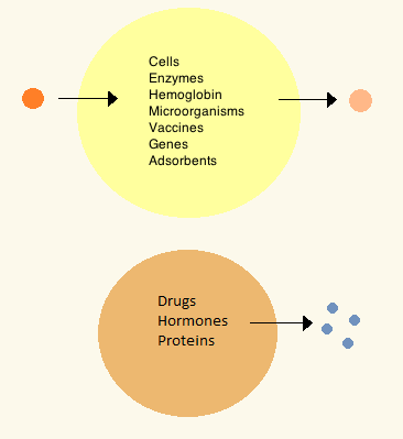
ในทศวรรษ 1960 โทมัส ชางได้พัฒนาไมโครแคปซูล ซึ่งต่อมาเขาจะเรียกว่า "เซลล์เทียม" เนื่องจากเป็นช่องขนาดเท่าเซลล์ที่ทำจากวัสดุเทียม[ 31 ]เซลล์เหล่านี้ประกอบด้วยเยื่อบางพิเศษที่ทำจากไนลอน คอลโลเดียน หรือโปรตีนที่เชื่อมโยงกัน ซึ่งคุณสมบัติกึ่งซึมผ่านได้ ทำให้โมเลกุล ขนาด เล็กสามารถ แพร่เข้าและออกจากเซลล์ได้ เซลล์เหล่านี้มีขนาดไมครอนและประกอบด้วยเซลล์เอนไซม์ฮี โม โกลบินวัสดุแม่เหล็ก สารดูดซับและโปรตีน [ 3 ]
ต่อมาเซลล์เทียมมีขนาดตั้งแต่ร้อยไมโครเมตรไปจนถึงนาโนเมตร และสามารถบรรจุจุลินทรีย์วัคซีนยีนยาฮอร์โมนและเปปไทด์ได้[ 3 ] การใช้เซลล์เทียมในทางคลินิกครั้งแรกคือการกรองเลือดโดยการห่อหุ้มถ่านกัมมันต์[ 32 ]
In the 1970s, researchers were able to introduce enzymes, proteins and hormones to biodegradable microcapsules, later leading to clinical use in diseases such as Lesch–Nyhan syndrome.[33] Although Chang's initial research focused on artificial red blood cells, only in the mid-1990s were biodegradable artificial red blood cells developed.[34] Artificial cells in biological cell encapsulation were first used in the clinic in 1994 for treatment in a diabetic patient[35] and since then other types of cells such as hepatocytes, adult stem cells and genetically engineered cells have been encapsulated and are under study for use in tissue regeneration.[36][37]
Materials

Membranes for artificial cells can be made of simple polymers, crosslinked proteins, lipid membranes or polymer-lipid complexes. Further, membranes can be engineered to present surface proteins such as albumin, antigens, Na/K-ATPase carriers, or pores such as ion channels. Commonly used materials for the production of membranes include hydrogel polymers such as alginate, cellulose and thermoplastic polymers such as hydroxyethyl methacrylate-methyl methacrylate (HEMA- MMA), polyacrylonitrile-polyvinyl chloride (PAN-PVC), as well as variations of the above-mentioned.[4] The material used determines the permeability of the cell membrane, which depends on the polymer choice and is important for determining adequate diffusion of nutrients, waste and other critical molecules. Hydrophilic polymers have the potential to be biocompatible and can be fabricated into a variety of forms which include polymer micelles, sol-gel mixtures, physical blends and crosslinked particles and nanoparticles.[4] Of special interest are stimuli-responsive polymers that respond to pH or temperature changes for the use in targeted delivery. These polymers may be administered in the liquid form through a macroscopic injection and solidify or gel in situ because of the difference in pH or temperature. Nanoparticle and liposome preparations are also routinely used for material encapsulation and delivery. A major advantage of liposomes is their ability to fuse to cell and organelle membranes.
Preparation
Many variations for artificial cell preparation and encapsulation have been developed. Typically, vesicles such as a nanoparticle, polymersome or liposome are synthesized. An emulsion is typically made through the use of high pressure equipment such as a high pressure homogenizer or a Microfluidizer. Two micro-encapsulation methods for nitrocellulose are also described below.
High-pressure homogenization
In a high-pressure homogenizer, two liquids in oil/liquid suspension are forced through a small orifice under very high pressure. This process divides the products and allows the creation of extremely fine particles, as small as 1 nm.
Microfluidization
This technique uses a patented Microfluidizer to obtain a greater amount of homogenous suspensions that can create smaller particles than homogenizers. A homogenizer is first used to create a coarse suspension which is then pumped into the microfluidizer under high pressure. The flow is then split into two streams which will react at very high velocities in an interaction chamber until desired particle size is obtained.[38] This technique allows for large scale production of phospholipid liposomes and subsequent material nanoencapsulations.
Drop method
In this method, a cell solution is incorporated dropwise into a collodion solution of cellulose nitrate. As the drop travels through the collodion, it is coated with a membrane due to the interfacial polymerization properties of the collodion. The cell later settles into paraffin, where the membrane sets, which is then suspended using a saline solution. The drop method is used for the creation of large artificial cells which encapsulate biological cells, stem cells and genetically engineered stem cells.
Emulsion method
The emulsion method differs in that the material to be encapsulated is usually smaller and is placed in the bottom of a reaction chamber where the collodion is added on top and centrifuged, or otherwise disturbed in order to create an emulsion. The encapsulated material is then dispersed and suspended in saline solution.
Clinical relevance
Drug release and delivery
Artificial cells used for drug delivery differ from other artificial cells since their contents are intended to diffuse out of the membrane, or be engulfed and digested by a host target cell. Often used are submicron, lipid membrane artificial cells that may be referred to as nanocapsules, nanoparticles, polymersomes, or other variations of the term.[39]
A temperature-responsive system has been developed to use RNA thermometers to control the timing and location of cargo release from artificial cells.[40] This is done by having artificial cells express a pore forming protein - alpha hemolysin - under the control of an RNA thermometer, allowing for cargo release to be coupled to temperature changes.[40]
Enzyme therapy
Enzyme therapy is being actively studied for genetic metabolic diseases where an enzyme is over-expressed, under-expressed, defective, or completely absent. In the case of under-expression or expression of a defective enzyme, an active form of the enzyme is introduced in the body to compensate for the deficit. On the other hand, an enzymatic over-expression may be counteracted by introduction of a competing non-functional enzyme; that is, an enzyme which metabolizes the substrate into non-active products. When placed within an artificial cell, enzymes can carry out their function for a much longer period compared to free enzymes[3] and can be further optimized by polymer conjugation.[41]
The first enzyme studied under artificial cell encapsulation was asparaginase for the treatment of lymphosarcoma in mice. This treatment delayed the onset and growth of the tumor.[42] These initial findings led to further research in the use of artificial cells for enzyme delivery in tyrosine dependent melanomas.[43] These tumors have a higher dependency on tyrosine than normal cells for growth, and research has shown that lowering systemic levels of tyrosine in mice can inhibit growth of melanomas.[44] The use of artificial cells in the delivery of tyrosinase; and enzyme that digests tyrosine, allows for better enzyme stability and is shown effective in the removal of tyrosine without the severe side-effects associated with tyrosine deprivation in the diet.[45]
Artificial cell enzyme therapy is also of interest for the activation of prodrugs such as ifosfamide in certain cancers. Artificial cells encapsulating the cytochrome p450 enzyme which converts this prodrug into the active drug can be tailored to accumulate in the pancreatic carcinoma or implanting the artificial cells close to the tumor site. Here, the local concentration of the activated ifosfamide will be much higher than in the rest of the body thus preventing systemic toxicity.[46] The treatment was successful in animals[47] and showed a doubling in median survivals amongst patients with advanced-stage pancreatic cancer in phase I/II clinical trials, and a tripling in one-year survival rate.[46]
Gene therapy
ในการรักษาโรคทางพันธุกรรมการบำบัดด้วยยีนมีเป้าหมายเพื่อแทรก เปลี่ยนแปลง หรือกำจัดยีนภายในเซลล์ของผู้ป่วย เทคโนโลยีนี้อาศัยเวกเตอร์ ไวรัสเป็นอย่างมาก ซึ่งก่อให้เกิดความกังวลเกี่ยวกับการกลายพันธุ์ จากการแทรก และการตอบสนองทางภูมิคุ้มกัน ทั่วร่างกาย ซึ่งนำไปสู่การเสียชีวิตของมนุษย์[ 48 ] [ 49 ]และการเกิดโรคมะเร็งเม็ดเลือดขาว[ 50 ] [ 51 ]ในการทดลองทางคลินิก การหลีกเลี่ยงความจำเป็นในการใช้เวกเตอร์โดยใช้ดีเอ็นเอเปล่าหรือพลาสมิดเป็นระบบนำส่งเองก็พบปัญหาเช่นกัน เช่น ประสิทธิภาพ การถ่ายทอด ต่ำ และการกำหนดเป้าหมายเนื้อเยื่อที่ไม่ดีเมื่อให้ทางระบบ[ 4 ]
เซลล์เทียมได้รับการเสนอให้เป็นเวกเตอร์ที่ไม่ใช่ไวรัส โดยเซลล์ที่ไม่ใช่เซลล์ออโตโลกัสที่ได้รับการดัดแปลงทางพันธุกรรมจะถูกห่อหุ้มและปลูกถ่ายเพื่อส่งโปรตีนรีคอมบิแนนท์ในร่างกาย[ 52 ]การแยกภูมิคุ้มกันประเภทนี้ ได้รับการพิสูจน์แล้วว่ามีประสิทธิภาพในหนู โดยการส่งเซลล์เทียมที่มีฮอร์โมนการเจริญเติบโตของ หนู ซึ่งช่วยฟื้นฟูการเจริญเติบโตที่ช้าลงในหนูที่กลายพันธุ์[ 53 ]กลยุทธ์บางอย่างได้ก้าวหน้าไปสู่การทดลองทางคลินิกในมนุษย์สำหรับการรักษาโรคมะเร็งตับอ่อนโรคกล้ามเนื้ออ่อนแรง และการควบคุมความเจ็บปวด[ 4 ]
การฟอกเลือด
การใช้เซลล์เทียมในทางคลินิกครั้งแรกคือการฟอกเลือดโดยการห่อหุ้มถ่านกัมมันต์ [ 32 ] ถ่านกัมมันต์มีความสามารถในการดูดซับโมเลกุลขนาดใหญ่จำนวนมาก และเป็นที่รู้จักกันมานานแล้วว่ามีความสามารถในการกำจัดสารพิษออกจากเลือดในกรณีที่ได้รับพิษโดยไม่ได้ตั้งใจหรือได้รับยาเกินขนาด อย่างไรก็ตามการฟอกเลือดโดยการให้ถ่านกัมมันต์โดยตรงนั้นเป็นพิษ เนื่องจากทำให้เกิด ลิ่มเลือด อุดตันและความเสียหายของเซลล์เม็ดเลือดตามมาด้วยการกำจัดโดยเกล็ดเลือด[ 54 ] เซลล์เทียมช่วยให้สารพิษแพร่กระจายเข้าไปในเซลล์ได้ ในขณะที่กักเก็บสารอันตรายไว้ภายในเยื่อหุ้มเซลล์ที่บางมาก[ 32 ]
การกรอง เลือด ด้วยเซลล์เทียมได้รับการเสนอให้เป็นทางเลือกในการล้างพิษที่มีต้นทุนต่ำกว่าและมีประสิทธิภาพมากกว่าการฟอกไต [ 3 ] ซึ่งการกรองเลือดเกิดขึ้นโดยการแยกขนาดด้วยเยื่อทางกายภาพเท่านั้น ใน การกรองเลือด เซลล์เทียมดูดซับหลายพันเซลล์จะถูกกักเก็บไว้ภายในภาชนะขนาดเล็กโดยใช้ตะแกรงสองอันที่ปลายทั้งสองข้างซึ่งเลือดของผู้ป่วยจะไหลผ่านเมื่อเลือดไหลเวียนสารพิษหรือยาจะแพร่เข้าสู่เซลล์และถูกกักเก็บไว้โดยวัสดุดูดซับ เยื่อของเซลล์เทียมนั้นบางกว่าเยื่อที่ใช้ในการฟอกไตมาก และขนาดที่เล็กหมายความว่ามีพื้นที่ผิว ของเยื่อสูง ซึ่งหมายความว่าส่วนหนึ่งของเซลล์สามารถมีการถ่ายโอนมวลตามทฤษฎีได้สูงกว่าเครื่องไตเทียมทั้งเครื่องถึงร้อยเท่า[ 3 ]อุปกรณ์นี้ได้รับการยอมรับว่าเป็นวิธีการทางคลินิกประจำสำหรับผู้ป่วยที่ได้รับการรักษาจากการได้รับสารพิษโดยอุบัติเหตุหรือการฆ่าตัวตาย แต่ยังได้รับการแนะนำให้ใช้เป็นวิธีการรักษาในภาวะตับวายและไตวายโดยทำหน้าที่บางส่วนของอวัยวะเหล่านี้[ 3 ] การฟอกเลือดด้วยเซลล์เทียมยังได้รับการเสนอให้ใช้ในการดูดซับภูมิคุ้มกัน ซึ่งสามารถกำจัดแอนติบอดีออกจากร่างกายได้โดยการติดวัสดุดูดซับภูมิคุ้มกัน เช่นอัลบูมินบนพื้นผิวของเซลล์เทียม หลักการนี้ถูกนำมาใช้เพื่อกำจัด แอนติบอดี หมู่เลือดออกจากพลาสมาสำหรับการปลูกถ่ายไขกระดูก[ 55 ]และสำหรับการรักษาภาวะคอเลสเตอรอลสูงผ่านแอนติบอดีโมโนโคลนอล เพื่อกำจัด ไลโปโปรตีนความหนาแน่นต่ำ[ 56 ]การฟอกเลือดมีประโยชน์อย่างยิ่งในประเทศที่มีอุตสาหกรรมการผลิตเครื่องฟอกไตที่อ่อนแอ เนื่องจากอุปกรณ์เหล่านี้มักมีราคาถูกกว่าและใช้กับผู้ป่วย โรคไตวาย
เซลล์ที่ถูกห่อหุ้ม

The most common method of preparation of artificial cells is through cell encapsulation. Encapsulated cells are typically achieved through the generation of controlled-size droplets from a liquid cell suspension which are then rapidly solidified or gelled to provide added stability. The stabilization may be achieved through a change in temperature or via material crosslinking.[4] The microenvironment that a cell sees changes upon encapsulation. It typically goes from being on a monolayer to a suspension in a polymer scaffold within a polymeric membrane. A drawback of the technique is that encapsulating a cell decreases its viability and ability to proliferate and differentiate.[57] Further, after some time within the microcapsule, cells form clusters that inhibit the exchange of oxygen and metabolic waste,[58] leading to apoptosis and necrosis thus limiting the efficacy of the cells and activating the host's immune system. Artificial cells have been successful for transplanting a number of cells including islets of Langerhans for diabetes treatment,[59]parathyroid cells and adrenal cortex cells.
Encapsulated hepatocytes
Shortage of organ donors make artificial cells key players in alternative therapies for liver failure. The use of artificial cells for hepatocyte transplantation has demonstrated feasibility and efficacy in providing liver function in models of animal liver disease and bioartificial liver devices.[60] Research stemmed off experiments in which the hepatocytes were attached to the surface of a micro-carriers[61] and has evolved into hepatocytes which are encapsulated in a three-dimensional matrix in alginate microdroplets covered by an outer skin of polylysine. A key advantage to this delivery method is the circumvention of immunosuppression therapy for the duration of the treatment. Hepatocyte encapsulations have been proposed for use in a bioartificial liver. The device consists of a cylindrical chamber imbedded with isolated hepatocytes through which patient plasma is circulated extra-corporeally in a type of hemoperfusion. Because microcapsules have a high surface area to volume ratio, they provide large surface for substrate diffusion and can accommodate a large number of hepatocytes. Treatment to induced liver failure mice showed a significant increase in the rate of survival.[60] Artificial liver systems are still in early development but show potential for patients waiting for organ transplant or while a patient's own liver regenerates sufficiently to resume normal function. So far, clinical trials using artificial liver systems and hepatocyte transplantation in end-stage liver diseases have shown improvement of health markers but have not yet improved survival.[62] The short longevity and aggregation of artificial hepatocytes after transplantation are the main obstacles encountered. Hepatocytes co-encapsulated with stem cells show greater viability in culture and after implantation[63] and implantation of artificial stem cells alone have also shown liver regeneration.[64] As such interest has arisen in the use of stem cells for encapsulation in regenerative medicine.
Encapsulated bacterial cells
The oral ingestion of live bacterial cell colonies has been proposed and is currently in therapy for the modulation of intestinal microflora,[65] prevention of diarrheal diseases,[66] treatment of H. Pylori infections, atopic inflammations,[67]lactose intolerance[68] and immune modulation,[69] amongst others. The proposed mechanism of action is not fully understood but is believed to have two main effects. The first is the nutritional effect, in which the bacteria compete with toxin producing bacteria. The second is the sanitary effect, which stimulates resistance to colonization and stimulates immune response.[4] The oral delivery of bacterial cultures is often a problem because they are targeted by the immune system and often destroyed when taken orally. Artificial cells help address these issues by providing mimicry into the body and selective or long term release thus increasing the viability of bacteria reaching the gastrointestinal system.[4] In addition, live bacterial cell encapsulation can be engineered to allow diffusion of small molecules including peptides into the body for therapeutic purposes.[4] Membranes that have proven successful for bacterial delivery include cellulose acetate and variants of alginate.[4] Additional uses that have arisen from encapsulation of bacterial cells include protection against challenge from M. Tuberculosis[70] and upregulation of Ig secreting cells from the immune system.[71] The technology is limited by the risk of systemic infections, adverse metabolic activities and the risk of gene transfer.[4] However, the greater challenge remains the delivery of sufficient viable bacteria to the site of interest.[4]
Artificial blood cells as oxygen carriers
Nano sized oxygen carriers are used as a type of red blood cell substitutes, although they lack other components of red blood cells. They are composed of a synthetic polymersome or an artificial membrane surrounding purified animal, human or recombinant hemoglobin.[72] Overall, hemoglobin delivery continues to be a challenge because it is highly toxic when delivered without any modifications. In some clinical trials, vasopressor effects have been observed.[73][74]
Artificial red blood cells
Research interest in the use of artificial cells for blood arose after the AIDS scare of the 1980s. Besides bypassing the potential for disease transmission, artificial red blood cells are desired because they eliminate drawbacks associated with allogenic blood transfusions such as blood typing, immune reactions and its short storage life of 42 days. A hemoglobin substitute may be stored at room temperature and not under refrigeration for more than a year.[3] Attempts have been made to develop a complete working red blood cell which comprises carbonic not only an oxygen carrier but also the enzymes associated with the cell. The first attempt was made in 1957 by replacing the red blood cell membrane by an ultrathin polymeric membrane[75] which was followed by encapsulation through a lipid membrane[76] and more recently a biodegradable polymeric membrane.[3] A biological red blood cell membrane including lipids and associated proteins can also be used to encapsulate nanoparticles and increase residence time in vivo by bypassing macrophage uptake and systemic clearance.[77]
Artificial leuko-polymersomes
A leuko-polymersome is a polymersome engineered to have the adhesive properties of a leukocyte.[78] Polymersomes are vesicles composed of a bilayer sheet that can encapsulate many active molecules such as drugs or enzymes. By adding the adhesive properties of a leukocyte to their membranes, they can be made to slow down, or roll along epithelial walls within the quickly flowing circulatory system.
Unconventional types of artificial cells
Electronic artificial cell
The concept of an Electronic Artificial Cell has been expanded in a series of 3 EU projects coordinated by John McCaskill from 2004 to 2015.
คณะกรรมาธิการยุโรปสนับสนุนการพัฒนาโครงการวิวัฒนาการเซลล์เทียมที่ตั้งโปรแกรมได้ (PACE) [ 79 ]ตั้งแต่ปี 2004 ถึง 2008 โดยมีเป้าหมายเพื่อวางรากฐานสำหรับการสร้าง "หน่วยอิสระขนาดเล็กที่จัดระเบียบตัวเอง จำลองตัวเอง และวิวัฒนาการได้ ซึ่งสร้างขึ้นจากสารอินทรีย์และอนินทรีย์อย่างง่ายที่สามารถตั้งโปรแกรมทางพันธุกรรมเพื่อทำหน้าที่เฉพาะ" [ 79 ]เพื่อการบูรณาการเข้ากับระบบสารสนเทศในที่สุด โครงการ PACE ได้พัฒนา Omega Machine เครื่องแรก[ 80 ]ซึ่งเป็นระบบสนับสนุนชีวิตแบบไมโครฟลูอิดิกสำหรับเซลล์เทียมที่สามารถเสริมฟังก์ชันการทำงานที่ขาดหายไปทางเคมี (ตามที่ Norman Packard, Steen Rasmussen, Mark Beadau และ John McCaskill เสนอไว้แต่เดิม) เป้าหมายสูงสุดคือการบรรลุเซลล์ไฮบริดที่วิวัฒนาการได้ในสภาพแวดล้อมที่ตั้งโปรแกรมได้ระดับไมโครที่ซับซ้อน จากนั้นฟังก์ชันของ Omega Machine สามารถถูกลบออกทีละขั้นตอน ซึ่งก่อให้เกิดความท้าทายด้านวิวัฒนาการที่สามารถแก้ไขได้หลายประการต่อเคมีของเซลล์เทียม โครงการนี้ประสบความสำเร็จในการบูรณาการทางเคมีจนถึงระดับคู่ของฟังก์ชันหลักสามประการของเซลล์เทียม (ระบบย่อยทางพันธุกรรม ระบบกักเก็บ และระบบเมตาบอลิซึม) และสร้างสภาพแวดล้อมไมโครฟลูอิดิกแบบตั้งโปรแกรมได้ที่มีความละเอียดเชิงพื้นที่แบบใหม่สำหรับการบูรณาการการกักเก็บและการขยายพันธุกรรม[ 79 ] โครงการนี้นำไปสู่การสร้างศูนย์เทคโนโลยีสิ่งมีชีวิตแห่งยุโรป[ 81 ]
จากการวิจัยนี้ ในปี 2550 จอห์น แมคแคสคิลล์ ได้เสนอให้มุ่งเน้นไปที่เซลล์เทียมที่เสริมด้วยระบบอิเล็กทรอนิกส์ ซึ่งเรียกว่า เซลล์เคมีอิเล็กทรอนิกส์ (Electronic Chemical Cell) แนวคิดหลักคือการใช้แถวอิเล็กโทรดแบบขนานจำนวนมากที่เชื่อมต่อกับวงจรอิเล็กทรอนิกส์เฉพาะที่ในฟิล์มบางสองมิติ เพื่อเสริมการทำงานของเซลล์เคมีที่กำลังพัฒนาขึ้น ข้อมูลอิเล็กทรอนิกส์เฉพาะที่ซึ่งกำหนดวงจรการสลับและการตรวจจับของอิเล็กโทรดสามารถทำหน้าที่เป็นจีโนมอิเล็กทรอนิกส์ เสริมข้อมูลลำดับโมเลกุลในโปรโตคอลที่กำลังเกิดขึ้นใหม่ ข้อเสนอการวิจัยประสบความสำเร็จกับคณะกรรมาธิการยุโรปและทีมงานนักวิทยาศาสตร์นานาชาติซึ่งบางส่วนทับซ้อนกับกลุ่ม PACE ได้เริ่มทำงานในโครงการเซลล์เคมีอิเล็กทรอนิกส์ระหว่างปี 2551-2555 โครงการนี้แสดงให้เห็นว่า การขนส่งเฉพาะที่ที่ควบคุมด้วยระบบอิเล็กทรอนิกส์ของลำดับเฉพาะสามารถใช้เป็นระบบควบคุมเชิงพื้นที่เทียมสำหรับการแพร่กระจายทางพันธุกรรมของเซลล์เทียมในอนาคต และกระบวนการหลักของการเผาผลาญสามารถส่งผ่านได้โดยแถวอิเล็กโทรดที่เคลือบอย่างเหมาะสม
ข้อจำกัดที่สำคัญของแนวทางนี้ นอกเหนือจากความยากลำบากในเบื้องต้นในการควบคุมเคมีไฟฟ้าและจลนไฟฟ้าในระดับจุลภาคแล้ว คือระบบอิเล็กทรอนิกส์เชื่อมต่อกันเป็นชิ้นส่วนฮาร์ดแวร์ขนาดใหญ่ที่ไม่เป็นอิสระและแข็งตัว ในปี 2011 McCaskill เสนอให้กลับด้านเรขาคณิตของอิเล็กทรอนิกส์และเคมี กล่าวคือ แทนที่จะวางสารเคมีในตัวกลางอิเล็กทรอนิกส์ที่ทำงานอยู่ ให้วางอิเล็กทรอนิกส์ขนาดเล็กที่เป็นอิสระในตัวกลางทางเคมี เขาได้จัดโครงการเพื่อจัดการกับเซลล์เทียมอิเล็กทรอนิกส์รุ่นที่สามที่ระดับ 100 ไมโครเมตร ซึ่งสามารถประกอบตัวเองจาก "แผ่นเซลล์" สองแผ่นเพื่อล้อมรอบพื้นที่ทางเคมีภายใน และทำงานโดยอาศัยอิเล็กทรอนิกส์ที่ทำงานอยู่ซึ่งขับเคลื่อนด้วยตัวกลางที่พวกมันจุ่มอยู่ เซลล์ดังกล่าวสามารถคัดลอกทั้งเนื้อหาอิเล็กทรอนิกส์และเคมี และจะสามารถวิวัฒนาการได้ภายในข้อจำกัดที่กำหนดโดยบล็อกตัวสร้างขนาดเล็กที่สังเคราะห์ไว้ล่วงหน้าเป็นพิเศษ ในเดือนกันยายน 2012 งานในโครงการนี้ได้เริ่มต้นขึ้น[ 82 ]
เซลล์ประสาทเทียม
มีการวิจัยและพัฒนาเกี่ยวกับเซลล์ประสาทเทียมทางกายภาพ ทั้งแบบอินทรีย์และอนินทรีย์
ตัวอย่างเช่น เซลล์ประสาทเทียมบางชนิดสามารถรับ[ 83 ] [ 84 ]และปล่อยโดปามีน ( สัญญาณเคมีแทนที่จะเป็นสัญญาณไฟฟ้า) และสื่อสารกับเซลล์กล้ามเนื้อและ สมองของหนูตามธรรมชาติ ซึ่งมีศักยภาพในการใช้งานในBCI / อุปกรณ์เทียม[ 85 ] [ 86 ]
เมมริสเตอร์ที่เข้ากันได้ทางชีวภาพและใช้พลังงานต่ำอาจช่วยให้สามารถสร้างเซลล์ประสาทเทียมซึ่งทำงานที่แรงดันไฟฟ้าของศักยภาพการกระทำ ทางชีวภาพ และสามารถใช้ประมวลผลสัญญาณการตรวจจับ ทางชีวภาพโดยตรง สำหรับการคำนวณแบบนิวโรโมฟิกและ/หรือการสื่อสารโดยตรงกับเซลล์ประสาททางชีวภาพ[ 87 ] [ 88 ] [ 89 ]
วงจรนิวโรโมฟิกอินทรีย์ที่ทำจากพอลิเมอร์เคลือบด้วยเจลที่มีไอออนสูงเพื่อให้วัสดุสามารถนำประจุไฟฟ้าได้เหมือนเซลล์ประสาทจริงได้ถูกสร้างขึ้นในหุ่นยนต์ ทำให้หุ่นยนต์สามารถเรียนรู้การรับรู้และการเคลื่อนไหวในโลกแห่งความเป็นจริง แทนที่จะเรียนรู้ผ่านการจำลองหรือโลกเสมือนจริง[ 90 ] [ 91 ]ยิ่งไปกว่านั้น เซลล์ประสาทสไปค์เทียมที่ทำจากสสารอ่อน (พอลิเมอร์) สามารถทำงานในสภาพแวดล้อมที่เกี่ยวข้องทางชีววิทยาและช่วยให้เกิดการสื่อสารแบบประสานงานระหว่างโดเมนเทียมและโดเมนทางชีววิทยา[ 92 ] [ 93 ]
จีวานู
Jeewanu protocells are synthetic chemical particles that possess cell-like structure and seem to have some functional living properties.[94] First synthesized in 1963 from simple minerals and basic organics while exposed to sunlight, it is still reported to have some metabolic capabilities, the presence of semipermeable membrane, amino acids, phospholipids, carbohydrates and RNA-like molecules.[94] However, the nature and properties of the Jeewanu remains to be clarified.[94][95]
Semi-artificial cyborg cells
การผสมผสานระหว่างชีววิทยาเชิงสังเคราะห์นาโนเทคโนโลยีและวิทยาศาสตร์วัสดุได้ถูกนำมาใช้เพื่อสร้างเซลล์ไซบอร์กแบคทีเรียหลายรูปแบบ[ 96 ] [ 97 ] [ 98 ]แบคทีเรียที่ได้รับการปรับปรุงทางกลประเภทต่างๆ เหล่านี้ถูกสร้างขึ้นด้วยหลักการผลิตแบบไบโอนิกที่ผสมผสานเซลล์ธรรมชาติเข้ากับวัสดุที่ไม่มีชีวิต ในปี 2548 นักวิจัยจากภาควิชาวิศวกรรมเคมี มหาวิทยาลัยเนบราสกา ลินคอล์นได้สร้างเซ็นเซอร์วัดความชื้นที่มีความไวสูงมากโดยการเคลือบแบคทีเรียBacillus cereusด้วยอนุภาคนาโนทองคำ ซึ่งเป็นครั้งแรกที่ใช้จุลินทรีย์ในการสร้างอุปกรณ์อิเล็กทรอนิกส์ และน่าจะเป็นแบคทีเรียไซบอร์กหรือวงจรเซลล์ไซบอร์กตัวแรก[ 99 ]นักวิจัยจากภาควิชาเคมี มหาวิทยาลัยแคลิฟอร์เนีย เบิร์กลีย์ได้ตีพิมพ์บทความหลายชุดในปี 2559 ซึ่งอธิบายถึงการพัฒนาแบคทีเรียไซบอร์กที่สามารถเก็บเกี่ยวแสงแดดได้อย่างมีประสิทธิภาพมากกว่าพืช[ 100 ]ในการศึกษาครั้งแรก นักวิจัยได้กระตุ้นการไวต่อแสงด้วยตนเองของแบคทีเรียที่ไม่สังเคราะห์แสงMoorella thermoaceticaด้วย อนุภาค นาโนแคดเมียมซัลไฟด์ ทำให้สามารถสังเคราะห์กรดอะซิติกจากคาร์บอนไดออกไซด์ได้[ 101 ]บทความติดตามผลได้อธิบายถึงการชี้แจงกลไกการถ่ายโอนอิเล็กตรอนจากสารกึ่งตัวนำไปยังแบคทีเรีย ซึ่งช่วยให้สามารถเปลี่ยนคาร์บอนไดออกไซด์และแสงแดดเป็นกรดอะซิติกได้[ 102 ]นักวิทยาศาสตร์จากภาควิชาวิศวกรรมชีวการแพทย์ มหาวิทยาลัยแคลิฟอร์เนีย เดวิสและAcademia Sinicaในไต้หวัน ได้พัฒนาแนวทางที่แตกต่างออกไปในการสร้างเซลล์ไซบอร์กโดยการประกอบไฮโดรเจลสังเคราะห์ภายในไซโตพลาส ซึม ของ เซลล์แบคทีเรีย Escherichia coliทำให้เซลล์เหล่านั้นไม่สามารถแบ่งตัวได้และทนต่อปัจจัยด้านสิ่งแวดล้อมยาปฏิชีวนะและความเครียดออกซิเดชันสูง[ 103 ]การฉีดไฮโดรเจล สังเคราะห์เข้าไปในเซลล์ทำให้เซลล์ไซบอร์กเหล่านี้มี โครงร่างเซลล์เทียม และความทนทานที่ได้รับทำให้เซลล์เหล่านี้อยู่ในตำแหน่งที่ดีที่จะกลายเป็นระบบ นำส่งยาประเภทใหม่ที่อยู่ระหว่างวัสดุสังเคราะห์แบบดั้งเดิมและระบบที่ใช้เซลล์
ดูเพิ่มเติม
สรุปเนื้อหา
ข้อมูลสำคัญจากบทความ
ข้อมูลสำคัญเกี่ยวกับ Artificial cell
An artificial cell, synthetic cell or minimal cell is an engineered particle that mimics one or many functions of a biological cell.
Bottom-up engineering of living artificial cells
German pathologist Rudolf Virchow proposed that not only does life arise from cells, but every cell comes from another cell; " Omnis cellula e cellula ".
Ethics and controversy
Protocell research has created controversy and opposing opinions, including critics of the vague definition of "artificial life". [ 15 ] The creation of a basic unit of life is the most pressing ethical concern.
International research community
In the mid-2010s the research community started recognising the need to unify the field of synthetic cell research, acknowledging that the task of constructing an entire living organism from non-living components was beyond the resources of a single country.