อ่าน 14 นาที
โรคฝีคัณฑสูตร
โรคฝีคัณฑสูตร เป็นโรคติดเชื้อที่ผิวหนังชนิดหนึ่ง ซึ่งมักเกิดขึ้นเป็นถุงน้ำ ระหว่างก้นทั้งสองข้าง และมักเกิดขึ้นที่ปลายด้านบน [ 1 ] [ 3 ] อาการอาจรวมถึงอาการปวด บวม และแดง [ 1 ]...
โรคฝีคัณฑสูตร
| โรคฝีคัณฑสูตร 123 | |
|---|---|
| ชื่ออื่นๆ | ถุงน้ำฝีที่ก้นกบ, ฝีหนองที่ก้นกบ, โพรงหนองที่ก้นกบ, ถุงน้ำ/ฝีคันที่กระดูกก้นกบ |
 | |
| โรคฝีหนองเฉียบพลันบริเวณร่องก้น ส่วนบน | |
| ความเชี่ยวชาญ | ศัลยกรรมทั่วไป , ศัลยกรรมลำไส้ใหญ่และทวารหนัก |
| อาการ | อาการปวด บวม แดง มีของเหลวไหลออกมา[ 1 ] |
| เริ่มต้นตามปกติ | วัยผู้ใหญ่ตอนต้น[ 2 ] |
| สาเหตุ | ขนคุดบริเวณร่องก้น |
| ปัจจัยเสี่ยง | โรคอ้วนประวัติครอบครัว ขนดก ( hirsutism ) ออกกำลังกายไม่เพียงพอ[ 2 ] |
| วิธีการวินิจฉัย | โดยพิจารณาจากอาการและการตรวจร่างกาย[ 2 ] |
| การวินิจฉัยแยกโรค | Hidradenitis suppurativa , ฝีในช่องท้อง , รูขุมขนอักเสบ[ 2 ] |
| การป้องกัน | การโกนบริเวณ[ 1 ] |
| การรักษา | การผ่าตัดและระบาย [ 2 ] การผ่าตัดเอาออก |
| ความถี่ | 3 ต่อ 10,000 ต่อปี[ 2 ] |
โรคฝีคัณฑสูตรเป็นโรคติดเชื้อที่ผิวหนังชนิดหนึ่ง ซึ่งมักเกิดขึ้นเป็นถุงน้ำระหว่างก้นทั้งสองข้างและมักเกิดขึ้นที่ปลายด้านบน[ 1 ] [ 3 ]อาการอาจรวมถึงอาการปวด บวม และแดง[ 1 ]อาจมีของเหลวไหลออกมาด้วย แต่พบไข้ได้น้อยมาก[ 1 ] [ 2 ]
ปัจจัยเสี่ยง ได้แก่โรคอ้วนประวัติครอบครัว การนั่งเป็นเวลานาน ปริมาณเส้นผมที่มากเกินไป และการออกกำลังกายไม่เพียงพอ[ 2 ]เชื่อกันว่ากลไกพื้นฐานเกี่ยวข้องกับกระบวนการทางกลที่เส้นผมและเศษผิวหนังถูกดูดเข้าไปในเนื้อเยื่อใต้ผิวหนังผ่านรูเปิดของผิวหนังที่เรียกว่าหลุม[ 2 ] การวินิจฉัยขึ้นอยู่กับอาการและการตรวจร่างกาย[ 2 ]
หากมีการติดเชื้อ การรักษาโดยทั่วไปคือการกรีดและระบายหนองบริเวณกึ่งกลาง[ 1 ] [ 2 ]การโกนขนบริเวณนั้นและการกำจัดขนด้วยเลเซอร์อาจช่วยป้องกันการกลับมาเป็นซ้ำได้[ 1 ] [ 4 ]อาจต้องผ่าตัดใหญ่ขึ้นหากโรคกลับมาเป็นซ้ำ[ 1 ] โดยปกติไม่จำเป็นต้องใช้ยาปฏิชีวนะ[ 2 ]หากไม่ได้รับการรักษา อาการอาจคงอยู่เป็นเวลานาน[ 1 ]
About 3 per 10,000 people per year are affected, and it occurs more often in males than females.[2] Young adults are most commonly affected.[2] The term pilonidal means 'nest of hair'.[1] The condition was first described in 1833.[1]
Signs and symptoms

Pilonidal cysts can be itchy and often very painful, and typically occur between the ages of 15 and 35.[5] Although usually found near the coccyx, the condition can also affect the navel, armpit, the cheek,[6] or the genital region,[7] though these locations are much rarer.
Signs and symptoms may include:[8]
- Intermittent pain/discomfort or swelling above the anus or near the tailbone
- Opaque yellow (purulent) or bloody discharge from the tailbone area
- Unexpected moisture in the tailbone region
- Discomfort sitting on the tailbone, doing sit-ups, or riding a bicycle—any activities that roll over the tailbone area
Some people with a pilonidal cyst will be asymptomatic.[9]
Pilonidal sinus
Pilonidal sinus (PNS): a sinus tract, or small channel, that may originate from the source of infection and open to the surface of the skin.[10] Material from the cyst drains through the pilonidal sinus. A pilonidal cyst is usually painful, but if it is a draining sinus, the pressure is relieved and the patient might not feel pain.
Causes
Hair insertion is the causative agent of pilonidal cysts.[11][12] An analysis of 624 patients' cyst hair found that 74% of the hair was rootless, and resembled spiky, razor-cut hair rather than intact body hair.[11] One proposed cause is ingrown hair,[13] although hairs found in pilonidal sinus tracts have more often been found to originate from the head.
Excessive sitting is thought to predispose people to the condition, as sitting increases pressure on the coccygeal region.
Trauma is not believed to cause a pilonidal cyst; however, such an event may result in inflammation of an existing cyst. There are cases where this has occurred months after a localized injury to the area.
ซีสต์พิโลนิดัลอาจเกิดจากรอยบุ๋มพิโลนิดัลแต่ กำเนิด [ 14 ]
เหงื่อออกมากเกินไปยังสามารถทำให้เกิดซีสต์ที่ก้นได้อีกด้วย ความชื้นสามารถเติมเต็มรูขุมขนที่ยืดออก ซึ่งช่วยสร้างสภาพแวดล้อมที่มีออกซิเจนต่ำซึ่งส่งเสริมการเจริญเติบโตของแบคทีเรียแบบไม่ใช้ออกซิเจน ซึ่งมักพบในซีสต์ที่ก้น การมีแบคทีเรียและระดับออกซิเจนต่ำจะขัดขวางการสมานแผลและทำให้ซีสต์ที่ก้นที่กำลังพัฒนาแย่ลง[ 15 ]
การวินิจฉัยแยกโรค

ซีสต์บริเวณร่องก้นอาจมีลักษณะคล้ายซีสต์เดอร์มอยด์ซึ่งเป็นเนื้องอกชนิดหนึ่ง ที่เรียกว่า เทอราโตมา ( เนื้องอกเซลล์สืบพันธุ์ ) โดยเฉพาะอย่างยิ่ง ซีสต์บริเวณร่อง ก้น อาจมีลักษณะคล้ายเทอราโตมาบริเวณกระดูกก้นกบการวินิจฉัยที่ถูกต้องมีความสำคัญ เนื่องจากเทอราโตมา ทุกชนิด จำเป็นต้องปรึกษาแพทย์ผู้เชี่ยวชาญด้านมะเร็งวิทยาและทำการผ่าตัดเอาออกทั้งหมด หากเป็นไปได้ควรหลีกเลี่ยงการรั่วไหลของเนื้อเยื่อ
การรักษา
ตามแนวทางปฏิบัติของสมาคมโรคลำไส้ใหญ่และทวารหนักแห่งยุโรป (ESCP) ปี 2024 ที่ตีพิมพ์ใน BJS เทคนิคการรักษา PSD ได้แก่ การระบายและการขูดการใช้ฟีนอล กาวไฟบริน การรักษาด้วยเลเซอร์การ ผ่าตัด เปิดช่องท้อง EPSiT การผ่าตัดแบบแผลเล็ก (MIS) การเจาะหนอง PRP และการตัดออกพร้อมการสมานแผลแบบเปิด[ 16 ]หากมีการติดเชื้อ การรักษาโดยทั่วไปคือการกรีดและระบายหนองออกจากแนวกึ่งกลาง เนื่องจากแผลที่แนวกึ่งกลางจะสมานแผลได้ยาก[ 1 ] [ 2 ]การปฏิบัติตามกฎง่ายๆ 5 ข้อเป็นที่ทราบกันดีว่าสามารถป้องกันการอักเสบซ้ำสำหรับบางคนและหลีกเลี่ยงการผ่าตัดได้: 1. หลีกเลี่ยงเก้าอี้และเบาะรถยนต์ที่กดทับกระดูกก้นกบ 2. มีน้ำหนักตัวอยู่ในเกณฑ์ปกติ โดยควรมีดัชนีมวลกาย (BMI) ต่ำ 3. รักษาบริเวณนั้นให้สะอาด 4. รักษาบริเวณนั้นให้แห้งโดยการสวมใส่เสื้อผ้าฝ้ายเท่านั้น 5. กำจัดขนบริเวณนั้นให้หมดจด เช่น โดยการใช้เครื่องกำจัดขนIPL เป็นประจำ [ 17 ]
The evidence for elective treatment of pilonidal sinus disease is poor.[18] The most commonly performed surgery is for the pilonidal sinus complex to be surgically excised with the wound often left open to heal. Post-surgical wound packing may be necessary, and packing typically must be replaced daily for four to eight weeks. In some cases, two years may be required for complete granulation to occur. Sometimes the cyst is resolved via surgical marsupialization.[19]
A 2018 literature review of 740 records of surgeries that included recurrence rates found that primary midline closure surgeries resulted in a 67.9% recurrence rate within 20 years, and recommended that they should be discontinued due to the high recurrence rate.[20] Incision and drainage had a recurrence rate of 25.9% within 2 years, up to 40.2% in 5 years. Phenol treatment has a recurrence rate of 14.1% at 2 years and 40.4% at 5 years.[20] A 2024 study involving 667 people found that, compared with tissue-removing surgery, minor procedures (such as draining and pit-picking) were associated with less pain, fewer complications and a faster recovery. However, minor surgeries were less likely to resolve the condition.[21][22]
Surgeons can also excise the sinus and repair it with a reconstructive flap technique, such as a "cleft lift" procedure or Z-plasty, usually done under general anesthetic. This approach is especially useful for complicated or recurring pilonidal disease, leaves little scar tissue, and flattens the region between the buttocks, reducing the risk of recurrence.[15] This approach typically results in a more rapid recovery than traditional surgery; however, there are fewer surgeons trained in the cleft lift procedure, and it consequently may not be as accessible to patients, depending on their location. Meta-analysis shows recurrence rates were lower in open healing than with primary closure (RR 0.60, 95% CI 0.42 to 0.87), at the expense of healing time.[23] Pilonidal cysts can recur, and do so more frequently if the surgical wound is sutured in the midline, as opposed to away from the midline, which obliterates the natal cleft and removes the focus of shearing stress. An incision lateral to the intergluteal cleft is therefore preferred, especially given the poor healing of midline incisions in this region. Minimally invasive techniques with no wound and rapid return to full activities have been reported but await double-blind randomized trials.[24]
เทคนิคอีกอย่างหนึ่งคือการรักษาฝีที่ก้นด้วยกาวไฟบรินเทคนิคนี้มีประโยชน์ไม่ชัดเจน ณ ปี 2017 เนื่องจากการวิจัยยังไม่เพียงพอ[ 25 ]หลักฐานสำหรับการรักษาใดๆ ก็ตามมีคุณภาพต่ำ และต้องระมัดระวังไม่ให้ตีความการศึกษาใดๆ ในสาขานี้มากเกินไป[ 18 ]
นับตั้งแต่ทศวรรษ 2010 เป็นต้นมา มีการพัฒนาเทคนิคการผ่าตัดแบบแผลเล็กหลายวิธีเพื่อลดผลกระทบของการผ่าตัดต่อผู้ป่วยและลดความเจ็บปวดและลดระยะเวลาการฟื้นตัว[ 26 ]
ในบางกรณี แผลจะถูกปล่อยให้เปิดไว้หลังการผ่าตัดเพื่อให้หายเองตามธรรมชาติแทนที่จะเย็บปิด มีวัสดุปิดแผลและสารทาเฉพาะที่ (ครีมหรือโลชั่น) หลายชนิดที่ช่วยให้แผลเปิดเหล่านี้หายเร็วขึ้น การทบทวนอย่างเป็นระบบในปี 2022 ได้รวบรวมหลักฐานจาก 11 การศึกษาที่เปรียบเทียบวัสดุปิดแผลและสารทาเฉพาะที่สำหรับการรักษาแผลเปิดหลังการผ่าตัดรักษาโรคฝีคัณฑสูตรที่ก้น[ 27 ]ผู้เขียนสรุปว่า: พลาสมาที่อุดมไปด้วยเกล็ดเลือดอาจช่วยให้แผลหายเร็วขึ้นเมื่อเทียบกับผ้าก๊อซปลอดเชื้อ ครีมซ่อมแซมผิว Lietofix อาจช่วยให้แผลหายภายใน 30 วันเมื่อเทียบกับไอโอดีน (ซึ่งช่วยลดแบคทีเรียในแผล) แต่ยังไม่ชัดเจนว่า วัสดุปิดแผลไฮโดรเจล (ที่ออกแบบมาเพื่อรักษาความชุ่มชื้นของแผล) จะช่วยลดระยะเวลาในการรักษาแผลเมื่อเทียบกับการทำความสะอาดแผลด้วยไอโอดีนหรือไม่[ 27 ]
การรักษาฝีที่ก้นกบด้วยกล้องเอน โดสโคปซึ่งใช้กล้องขนาดเล็กนำทางศัลยแพทย์ในการกำจัดขน เป็นวิธีการรักษาแบบใหม่ที่มีความเจ็บปวดน้อยและหายเร็วเมื่อเทียบกับการผ่าตัด การทบทวนวรรณกรรมในผู้ป่วย 497 รายพบว่าเวลาเฉลี่ยในการผ่าตัดด้วยกล้องเอนโดสโคปคือ 34.7 นาที และเวลาในการหายเฉลี่ยคือ 32.9 วัน ความล้มเหลวเกิดขึ้นใน 8% ของผู้ป่วย ซึ่งมีโรคคงอยู่หรือกลับมาเป็นซ้ำ[ 28 ]
แนวทางปฏิบัติปัจจุบันของสมาคมศัลยกรรมลำไส้ใหญ่และทวารหนักแห่งยุโรป (ESCP) (2024) ระบุว่าเทคนิคที่ใช้เลเซอร์ เช่น การปิดไซนัสด้วยเลเซอร์ (SiLaC/SiLaT) อาจถือเป็น ทางเลือกการรักษาแบบ รุกรานน้อยที่สุดสำหรับผู้ป่วยบางรายที่เป็นโรคฝีคัณฑสูตร ซึ่งสะท้อนให้เห็นถึงฐานข้อมูลหลักฐานที่กำลังเกิดขึ้นใหม่แต่ยังคงพัฒนาต่อไป[ 29 ]
Several randomized controlled trials have compared SiLaC, SiLaT, or other laser ablation techniques with conventional surgical approaches such as excision, curettage, or flap procedures.[30][31][32] These studies, generally small and often single-center with short- to mid-term follow-up, report that laser-based techniques may be associated with reduced postoperative pain, shorter operative time, shorter hospital stay, faster wound healing, and improved cosmetic outcomes, with recurrence rates broadly comparable to conventional surgery in the short term.
Systematic reviews and meta-analyses of laser treatment in pilonidal disease report primary healing rates of approximately 80–85% and relatively low complication rates, with recurrence rates varying depending on follow-up duration and study design.[33][34] A broader meta-analysis of minimally invasive techniques suggests that laser ablation may be associated with a lower risk of recurrence compared with excisional surgery, although the included studies are heterogeneous.[35]
A systematic review by Romic et al. (2022), including 10 studies with 971 patients, reported a primary healing rate of 94.4% and a weighted mean recurrence rate of 3.8% following sinus laser-assisted closure.[36] The authors concluded that laser treatment represents a promising option for managing chronic PD based on the published literature.[37]
Overall, the available evidence suggests that laser-based techniques are a minimally invasive treatment option for pilonidal disease with favorable short-term outcomes, while long-term outcomes remain to be further evaluated, necessitating long-term studies to assess its effectiveness as a first-line treatment.
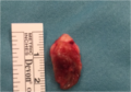 Excised pilonidal cyst
 Trephine/biopsy punch minimally invasive surgery for pilonidal disease (1)
 Trephine/biopsy punch minimally invasive surgery for pilonidal disease (2)
 Pilonidal cyst two days after traditional closed surgery
 Anatomy of pilonidal disease removed after trephine or biopsy punch surgery: pilonidal fistula (top) and pilonidal cyst (bottom)


Etymology
Pilonidal means 'nest of hair' and is derived from the Latin words for 'hair' (pilus) and 'nest' (nidus).[5] The condition was first described by Herbert Mayo in 1833.[38]R. M. Hodges was the first to use the phrase pilonidal cyst to describe the condition in 1880.[39][40]
The condition was widespread in the United States Army during World War II. The condition was termed "Jeep seat" or "Jeep riders' disease", because a large portion of people who were being hospitalized for it rode in Jeeps, and prolonged bumpy rides in the vehicles were believed to have caused the condition due to irritation and pressure on the coccyx.
External links
- NHS Choices for pilonidal sinus treatment
สรุปเนื้อหา
ข้อมูลสำคัญจากบทความ
ข้อมูลสำคัญเกี่ยวกับ โรคฝีคัณฑสูตร
โรคฝีคัณฑสูตร เป็นโรคติดเชื้อที่ผิวหนังชนิดหนึ่ง ซึ่งมักเกิดขึ้นเป็นถุงน้ำ ระหว่างก้นทั้งสองข้าง และมักเกิดขึ้นที่ปลายด้านบน [ 1 ] [ 3 ] อาการอาจรวมถึงอาการปวด บวม และแดง [ 1 ]...
Signs and symptoms
Pilonidal cysts can be itchy and often very painful, and typically occur between the ages of 15 and 35.
Pilonidal sinus
Pilonidal sinus (PNS): a sinus tract, or small channel, that may originate from the source of infection and open to the surface of the skin. [ 10 ] Material from the cyst drains through the pilonidal sinus.
Causes
Hair insertion is the causative agent of pilonidal cysts. [ 11 ] [ 12 ] An analysis of 624 patients' cyst hair found that 74% of the hair was rootless, and resembled spiky, razor-cut hair rather than intact body hair.