
Sacroiliac joint dysfunction
| Sacroiliac joint dysfunction | |
|---|---|
| Other names | Sacroiliac joint disorder, sacroiliac joint disease, sacroiliac joint syndrome, sacroiliac syndrome, sacroilliac dysfunction and instability |
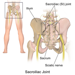 | |
| The sacroiliac joint, posterior view | |
The term sacroiliac joint dysfunction refers to abnormal motion in the sacroiliac joint, either too much motion or too little motion, that causes pain in this region.
Signs and symptoms
Common symptoms include lower back pain, buttocks pain, sciatic leg pain, groin pain, hip pain (for explanation of leg, groin, and hip pain, see referred pain), urinary frequency, and "transient numbness, prickling, or tingling".[1][2][3][4][5][6][7] Pain can range from dull aching to sharp and stabbing and increases with physical activity.[1][8][2] Symptoms also worsen with prolonged or sustained positions (i.e., sitting, standing, lying).[8][9][2] Bending forward, stair climbing, hill climbing, and rising from a seated position can also provoke pain. Pain can increase during menstruation in women.[8][9][2] People with severe and disabling sacroiliac joint dysfunction can develop insomnia and depression.[10] Sacral rotation can be transmitted distally down the kinematic chain and, if left untreated over a long period of time, may lead to severe Achilles tendinitis.[11]
Causes
Hypermobility
Sacroiliac joint dysfunction is an outcome of either extra-articular dysfunction or from intraarticular dysfunction.
ความผิดปกติของข้อต่อกระดูกเชิงกรานบางครั้งเรียกว่า "ความไม่เสถียรของข้อต่อกระดูกเชิงกราน" หรือ "ความไม่เพียงพอของข้อต่อกระดูกเชิงกราน" เนื่องจากเอ็นที่เคยแข็งแรงและตึงไม่สามารถรองรับได้อีกต่อไป[ 8 ] [ 9 ] [ 2 ] [ 12 ]เมื่อข้อต่อเคลื่อนไหวมากเกินไปหรือหลวม จะถูกจัดเป็นความผิดปกติภายนอกข้อต่อ เนื่องจากการเคลื่อนไหวและการจัดเรียงข้อต่อที่ผิดปกติเป็นผลมาจากเอ็นที่อ่อนแอ บาดเจ็บ หรือเคล็ดในขณะที่ข้อต่อเองมีโครงสร้างปกติและแข็งแรง ข้อต่อกระดูกเชิงกรานเองมักจะไม่แสดง การเปลี่ยนแปลง ที่เสื่อมสภาพเช่น โรคข้ออักเสบจนกว่าจะปล่อยให้ความผิดปกติดำเนินต่อไปเป็นเวลาหลายปี[ 8 ]การบาดเจ็บของเอ็นที่ยึดข้อต่อกระดูกเชิงกรานให้อยู่ในตำแหน่งที่เหมาะสมนั้นเชื่อว่าเกิดจากการบิดหรือการบาดเจ็บที่มีแรงกระแทกสูง (เช่น อุบัติเหตุทางรถยนต์) หรือการล้มอย่างแรง ส่งผลให้เกิดการเคลื่อนไหวมากเกินไป[ 8 ] มากถึง 58% ของผู้ที่ได้รับการวินิจฉัยว่าเป็นโรคปวดข้อกระดูกเชิงกรานมีสาเหตุมาจากการบาดเจ็บจากการตรวจร่างกาย[ 7 ] ข้อต่อที่เคยได้รับการยึดให้มั่นคงด้วยเอ็นที่แข็งแรง ตอนนี้ยืดออกมากเกินไป เคล็ดขัดยอก หรือฉีกขาด ทำให้เคลื่อนไหวเกินขอบเขตปกติ เชื่อกันว่าสิ่งนี้ส่งผลให้พื้นผิวกระดูกเชิงกรานและกระดูกสันหลังส่วนล่าง "ล็อก" ในลักษณะที่ไม่สอดคล้องกันหรือไม่สมมาตร (กระดูกเชิงกรานข้างหนึ่งเอียงไปข้างหน้า กระดูกเชิงกรานอีกข้างหนึ่งเอียงไปข้างหลัง) ทำให้เกิดอาการปวดที่อาจทำให้ทุพพลภาพได้[ 8 ]
ความไม่สมดุลของฮอร์โมน โดยเฉพาะอย่างยิ่งที่เกี่ยวข้องกับการตั้งครรภ์และฮอร์โมนรีแลกซินอาจทำให้เอ็นหย่อนยานส่งผลให้โครงสร้างกระดูกเชิงกรานอ่อนแอลง[ 13 ] ในระหว่างตั้งครรภ์ รีแลกซินทำหน้าที่เป็นกลไกตามธรรมชาติที่ช่วยให้กระดูกเชิงกรานของผู้หญิงขยายตัวเพื่อรองรับช่องคลอด[ 13 ]อาการปวดข้อเชิงกรานในสตรีหลังคลอดเชื่อว่าเกิดจากเอ็นที่ยืดออกไม่สามารถกลับคืนสู่ความตึงตัวตามปกติได้[ 13 ]สตรีที่คลอดบุตรตัวใหญ่หรือผู้ที่มีระยะเวลาการคลอดนานก็มีแนวโน้มที่จะเกิดอาการปวดข้อเชิงกรานเรื้อรังและไม่มั่นคงได้เช่นกัน
In some people, the sacroiliac joints reverse the normal concave-convex 'locking' relationship, which can lead to rotational misalignment.[8][9][2] The variation in joint configuration results in some sacroiliac joints being inherently weaker or more prone to misalignment.[9]
Certain biomechanical or muscle length imbalances may ultimately predispose a person to sacroiliac dysfunction and pain. Likely, this is a result of altered gait patterns and repetitive stress to the SI joint and related structures.[1] These conditions exist in persons with leg-length inequality, scoliosis, a history of polio, poor-quality footwear, and hip osteoarthritis.[1]
There is also a notable incidence of lumbar spinal fusion patients that present with sacroiliac pain and hypermobility, potentially due to the adjacent lumbar joints being fixed and unable to move. Clinical studies have found up to 75% of post-lumbar fusion patients develop SI joint degeneration within five years of surgery.[14]
Hypomobility
Pathological hypomobility (too little movement) of the sacroiliac joint is an intra-articular disorder in which the joint locks due to wearing down with age or degenerative joint disease.
Hypomobility of this kind can also occur with an inflammatory disease such as ankylosing spondylitis,[15]rheumatoid arthritis, or an infection.
Pathophysiology
The sacroiliac joint is a true diarthrodial joint that joins the sacrum to the pelvis.[1][8][16][17] The sacrum connects on the right and left sides to the ilia (pelvic bones) to form the sacroiliac joints. The pelvic girdle is made up of two innominate bones (the iliac bones) and the sacrum. The innominate bones join in the front of the pelvis to form the pubic symphysis, and at back of the sacrum to form the sacroiliac (SI) joints. Each innominate bone (ilium) joins the femur (thigh bone) to form the hip joint; thus the sacroiliac joint moves with walking and movement of the torso.[9]
ในข้อต่อนี้กระดูกอ่อนไฮอะลีนด้านกระดูกสันหลังส่วนล่างจะเคลื่อนที่ไปชนกับกระดูกอ่อนไฟโบรไมอัลเจียมด้านกระดูกเชิงกราน ข้อต่อกระดูกสันหลังส่วนล่างประกอบด้วยสันและร่องจำนวนมากที่ทำหน้าที่ในการรักษาเสถียรภาพ การศึกษาต่างๆ ได้บันทึกไว้ว่ามีการเคลื่อนไหวเกิดขึ้นที่ข้อต่อ ดังนั้นจึงอาจเกิดการเคลื่อนหลุดเล็กน้อยหรือแม้แต่ตำแหน่งที่ล็อกได้[ 8 ] [ 16 ] [ 2 ] [ 18 ] [ 3 ] [ 19 ] [ 12 ]
กล้ามเนื้อและเอ็นล้อมรอบและยึดติดกับข้อต่อ SI ทั้งด้านหน้าและด้านหลัง โดยส่วนใหญ่จะอยู่บนพื้นผิวของกระดูกเชิงกรานหรือกระดูกศักรัม สิ่งเหล่านี้ล้วนเป็นแหล่งที่มาของความเจ็บปวดและการอักเสบได้หากข้อต่อ SI ทำงานผิดปกติ[ 9 ] [ 2 ] ข้อต่อศักรัมและกระดูกเชิงกรานขึ้นอยู่กับโครงสร้างเอ็นที่แข็งแรงเพื่อการรองรับและความมั่นคง[ 9 ]เอ็นที่มักจะถูกรบกวนและ/หรือฉีกขาดมากที่สุดคือเอ็น iliolumbarและเอ็น sacroiliac ด้านหลัง[ 9 ]โครงสร้างเอ็นให้ความต้านทานต่อแรงเฉือนและแรงกด เอ็นด้านหน้าด้านหลัง และ ระหว่างกระดูก ที่อยู่ลึกจะต้านทานแรงกดของกระดูกศักรัมเมื่อเทียบกับกระดูกเชิงกราน[ 1 ] เอ็นที่อยู่ตื้นกว่า (เช่นเอ็น sacrotuberous ) จะตอบสนองต่อการเคลื่อนไหวแบบไดนามิก (เช่น การยกขาตรงระหว่างการเคลื่อนไหวทางกายภาพ) [ 1 ] The long dorsal sacroiliac ligament can become stretched in periods of increased lumbar lordosis (eg, during pregnancy ). [ 20 ]
กลุ่มกล้ามเนื้อที่ได้รับผลกระทบ
กล้ามเนื้อขนาดใหญ่และเล็กจำนวนมากมีความสัมพันธ์กับเอ็นของข้อต่อกระดูกเชิงกราน รวมถึงกล้ามเนื้อ พิริ ฟอร์มิส (ดู " กลุ่มอาการพิริฟอร์มิส " ซึ่งเป็นภาวะที่มักเกี่ยวข้องกับความผิดปกติของข้อต่อกระดูกเชิงกราน) กล้ามเนื้อเรคตัสเฟโม ริ ส กล้าม เนื้อกลูเตียส แม็กซิมั สและมินิมัสกล้าม เนื้ออี เรคเตอร์สไปเน กล้ามเนื้อแลทิสซิมัสดอ ร์ซี พังผืดทรวงอกและเอวและ กล้ามเนื้ออิลิอาคั ส[ 1 ] กล้ามเนื้อเหล่านี้อาจเกี่ยวข้องหรือหดเกร็งได้ทำให้เกิดอาการปวดและความผิดปกติของข้อต่อกระดูกเชิงกราน[ 1 ] [ 8 ] [ 2 ] [ 3 ] [ 19 ] [ 12 ] ข้อต่อกระดูกเชิงกรานเป็นโครงสร้างที่ไวต่อความเจ็บปวด มีเส้นประสาทจำนวนมากมาเลี้ยงโดยปลายประสาทอิสระที่ไม่มีปลอกไมอีลิน และ แขนงหลักด้านหลังของส่วนกระดูกสันหลัง L2-S3 ความเป็นไปได้ที่กว้างขวางของการรับเส้นประสาทอาจอธิบายได้ว่าทำไมความเจ็บปวดที่เกิดจากข้อต่อจึงสามารถแสดงออกมาได้หลายวิธี โดยมีรูปแบบการส่งต่อที่แตกต่างและเฉพาะเจาะจง (ดู " ความเจ็บปวดที่ส่งต่อ ") สำหรับผู้ป่วยแต่ละราย[ 1 ] [ 18 ]ผู้ป่วยที่มีความผิดปกติของข้อต่อกระดูกเชิงกรานยังสามารถเกิดอาการตึงและทำงานผิดปกติในกล้ามเนื้อแฮมสตริง ค วอดริเซปส์ เอ็นไอลิ โอทิเบียล (ดู " กลุ่มอาการเอ็นไอลิโอทิเบียล ") และกล้ามเนื้อสะโพกรวมถึงกล้ามเนื้อพีโซแอส บุคคลที่มีความผิดปกติของข้อต่อกระดูกเชิงกรานอย่างรุนแรงและเรื้อรังอาจเกิดภาวะกล้ามเนื้ออ่อนแรงและฝ่อทั่วร่างกายเนื่องจากข้อจำกัดของกิจกรรมและการออกกำลังกายที่ทำให้เกิดอาการปวดหลังส่วนล่าง[ 1 ]
การวินิจฉัย
Perhaps the biggest reason for misdiagnosis or lack of diagnosis of sacroiliac joint dysfunction is based on the inability of common radiological imaging to discern the disorder. Diagnostic testing, such as X-ray, CT scan, or MRI, do not usually reveal abnormalities; therefore, they cannot reliably be used for diagnosis of sacroiliac joint dysfunction.[9][2] There is a new imaging test SPECT/CT which can sometimes detect sacroiliac joint dysfunction. There is also a lack of evidence that sacroiliac joint mobility maneuvers (Gillet, Standing flexion test, and Seated Flexion test) detect motion abnormalities.[21][22] Given the inherent technical limitations of the visible and palpable signs from these sacroiliac joint mobility maneuvers another broad category of clinical signs have been described called provocative maneuvers. These maneuvers are designed to reproduce or increase pain emanating within the sacroiliac joint.
A clinician (i.e., a spine surgeon, orthopedic surgeon, sports medicine doctor, athletic trainer, medical massage therapist, physical therapist, physiatrist, osteopath or chiropractor) can develop a probable diagnosis of sacroiliac joint dysfunction by using a hands on approach through palpating the painful areas and performing the following provocative maneuvers below:
- Gaenslen test - This pain provocation test applies torsion to the joint. With one hip flexed onto the abdomen, the other leg is allowed to dangle off the edge of the table. Pressure should then be directed downward on the leg in order to achieve hip extension and stress the sacroiliac joint.[1][2]
- Iliac Gapping Test - Distraction can be performed to the anterior sacroiliac ligaments by applying pressure to the anterior superior iliac spine.[1]
- Iliac Compression Test - Apply compression to the joint with the patient lying on his or her side. Pressure is applied downward to the uppermost iliac crest.[1]
- FABER or Patrick test - To identify if pain may come from the sacroiliac joint during flexion, abduction, and external rotation, the clinician externally rotates the hip while the patient lies supine. Then, downward pressure is applied to the medial knee stressing both the hip and sacroiliac joint.[1][2][4]
- Thigh Thrust - This test applies anteroposterior shear stress on the SI joint. The patient lies supine with one hip flexed to 90 degrees. The examiner stands on the same side as the flexed leg. The examiner provides either a quick thrust or steadily increasing pressure through the line of the femur. The pelvis is stabilized at the sacrum or at the opposite ASIS with the hand of the examiner
Cautious interpretation is warranted because there are no biomechanical studies showing that the Thigh Thrust test isolates forces in the SIJ when performed at 90 degrees and due to intra-individual variation in body type, hip flexibility, general flexibility of the trunk and pelvis.[23] In all the tests, pain along the typical area raises suspicion for sacroiliac joint dysfunction. However no single test is very reliable in the diagnosis of sacroiliac joint dysfunction. It is important to remember true neurogenic weakness, numbness, or loss of reflex should alert the clinician to consider nerve root pathology.[24]
The current "gold standard" for diagnosis of sacroiliac joint dysfunction emanating within the joint is sacroiliac joint injection confirmed under fluoroscopy or CT-guidance using a local anesthetic solution. The diagnosis is confirmed when the patient reports a significant change in relief from pain and the diagnostic injection is performed on two separate visits. Published studies have used at least a 75 percent change in relief of pain before a response is considered positive and the sacroiliac joint deemed the source of pain.[25][26][27] However, several other injection studies have compared intra-articular with extra-articular injection, and indicate that the ligament injection behind the joint is oftentimes superior to injection in the joint and seems to be a very underutilized diagnostic tool.[24][28]
Misdiagnosis
In the early 1900s, dysfunction of the sacroiliac joint was a common diagnosis associated with low back and sciatic nerve pain.[18] However, research by Danforth and Wilson in 1925 concluded that the sacroiliac joint could not cause sciatic nerve pain because the joint does not have a canal in which the nerves can be entrapped against the joint.[29] The biomechanical relationship between the sacroiliac joint, the piriformis muscle (see "piriformis syndrome"), and the sciatic nerve had not yet been discovered.[18]
ในปี พ.ศ. 2477 ผลงานของ Mixter และ Barr ได้เปลี่ยนจุดเน้นในการวิจัยและการรักษาจากข้อต่อกระดูกเชิงกรานไปสู่หมอนรองกระดูกสันหลังเคลื่อนโดยเฉพาะหมอนรองกระดูกสันหลังส่วนเอว[ 30 ]การมุ่งเน้นทางการแพทย์เกี่ยวกับหมอนรองกระดูกเคลื่อนได้รับการพัฒนาเพิ่มเติมโดยการคิดค้นMRIในปี พ.ศ. 2520 [ 31 ]การวินิจฉัยและการให้ความสนใจกับหมอนรองกระดูกเคลื่อนมากเกินไป ทำให้ข้อต่อกระดูกเชิงกรานกลายเป็นแหล่งกำเนิดความเจ็บปวดที่ถูกมองข้ามในผู้ป่วยที่มีอาการปวดหลังส่วนล่างประมาณ 15% ถึง 25% [ 1 ] [ 8 ] [ 3 ] [ 5 ] [ 6 ] [ 7 ]
เอ็นในข้อต่อกระดูกเชิงกรานเป็นหนึ่งในเอ็นที่แข็งแรงที่สุดในร่างกาย และแพทย์หลายคนไม่คิดว่าจะมีโอกาสเกิดการเคล็ดหรือฉีกขาดได้ ความสงสัยเกี่ยวกับการมีอยู่ของความผิดปกติของข้อต่อกระดูกเชิงกรานในวงการแพทย์เพิ่มมากขึ้นจากการถกเถียงกันว่าข้อต่อกระดูกเชิงกรานเคลื่อนไหวมากน้อยเพียงใด มีรายงานความแตกต่างมากถึง 2–17 องศาในผลการตรวจทางคลินิก[ 32 ] [ 33 ] [ 34 ]
การรักษา
การรักษามักขึ้นอยู่กับระยะเวลาและความรุนแรงของอาการปวดและการทำงานผิดปกติ ในระยะเฉียบพลัน (1–2 สัปดาห์แรก) สำหรับอาการเคล็ดขัดยอกเล็กน้อยของข้อต่อกระดูกเชิงกราน ผู้ป่วยมักจะได้รับการแนะนำให้พักผ่อน ประคบเย็น/ร้อน จัดกระดูกสันหลัง[ 35 ]และกายภาพบำบัด นอกจากนี้ ยา ต้านการอักเสบก็อาจมีประโยชน์เช่นกัน[ 1 ] [ 4 ]
If the pain does not resolve in the first 1–2 weeks, then the patient may benefit from a steroid and anesthetic mixture fluoroscopically injected into the joint (this also serves in confirming diagnosis), as well as manipulative or manual therapy. [ 1 ] [ 8 ] [ 9 ] [ 2 ] [ 3 ] [ 12 ] For the most severe and chronic forms of sacroiliac dysfunction, treatment should proceed with the support of a sacroiliac belt, injection therapy, and finally, surgery . [ 1 ] [ 8 ] [ 9 ] [ 2 ] [ 18 ] The anti-inflammatory effect of injection therapy is not permanent, and the injections do not offer an opportunity to stabilize an incompetent joint. [ 36 ] Surgery is often considered a last resort, but for some patients, it is the only method of effectively stabilizing the loose joint. การตรึงข้อต่อ (โดยใช้สกรูหรืออุปกรณ์ที่คล้ายกันเท่านั้น โดยไม่ต้องใช้การปลูกถ่ายกระดูก ) เป็นที่นิยมมากกว่าการเชื่อมกระดูกสันหลังเนื่องจากเป็นการผ่าตัดที่รุกล้ำน้อยกว่า ตรงไปตรงมา และส่งผลให้ผู้ป่วยฟื้นตัวได้เร็วขึ้น[ 8 ] [ 9 ] [ 18 ] [ 37 ] ผู้เชี่ยวชาญบางคนในสาขานี้เชื่อว่าสิ่งสำคัญคือต้องแน่ใจว่าข้อต่อกระดูกเชิงกรานอยู่ในตำแหน่งที่ถูกต้องตามหลักกายวิภาคก่อนการตรึงหรือการเชื่อม แต่ผลการวิจัยที่ตีพิมพ์ขัดแย้งกับความเชื่อนี้[ 8 ] [ 9 ] [ 2 ] [ 18 ] [ 38 ] [ 39 ]
การฉีด พลาสม่าที่อุดมไปด้วยเกล็ดเลือด (PRP) แสดงให้เห็นผลลัพธ์เชิงบวกในการรักษาภาวะการทำงานผิดปกติของข้อต่อกระดูกเชิงกราน โดยการทดลองแบบสุ่มและรายงานกรณีศึกษาแสดงให้เห็นว่ามีประสิทธิภาพมากกว่าการฉีดสเตียรอยด์ในช่วงระยะเวลา 3 เดือน นอกจากนี้ การศึกษายังแสดงให้เห็นว่าการฉีด PRP เข้าไปในข้อต่อกระดูกเชิงกรานสามารถบรรเทาอาการได้อย่างสมบูรณ์และคงอยู่ได้นานถึงสี่ปี[ 40 ] [ 41 ] [ 42 ] [ 43 ]
Dextrose prolotherapy injections performed either intraarticularly or into the dorsal sacroiliac ligaments is sometimes performed as an alternative treatment option. This is a controversial therapy but it does have research evidence to support its use. Kim et al. in 2010 published a randomized controlled trial evaluating the effect of intraarticular prolotherapy injections versus intraarticular steroid injections in proven sacroiliac joint pain. The two interventions were equal in the short term at 3 months, but the prolotherapy group had superior pain relief at the 15 month mark at the end of follow up. This type of trial is considered high level evidence in medical circles.[44] Hoffman et al. in 2018 published a prospective cohort study with positive results in the treatment of those with sacroiliac joint dysfunction.[45]
Muscle energy techniques (MET) are commonly used as part of conservative treatment for sacroiliac joint dysfunction. MET is a manual therapy intervention in which the patient performs controlled muscle contractions against resistance provided by the clinician to improve joint mobility and reduce musculoskeletal dysfunction. This approach is frequently used to address pelvic asymmetry and restore normal lumbopelvic mechanics. Recent research has evaluated the effectiveness of MET in individuals with sacroiliac joint dysfunction. A 2024 study assessing the use of muscle energy techniques demonstrated significant improvements in pain and functional outcomes following treatment. Theses findings support the use of MET as an effective non-invasive intervention in the management of sacroiliac joint dysfunction. [11]
Exercise therapy for sacroiliac joint dysfunction typically focuses on improving lumbopelvic stability, strengthening core musculature, and enhancing neuromuscular control. Common approaches include strengthening of the gluteal muscles, abdominal stabilizers, and lumbar spine musculature, as well as stretching exercises targeting tight muscle groups around the pelvis and lower back. Exercises for the sacroiliac joint include sacroiliac joint self-mobilization, piriformis stretch, gluteus medius stretch, gluteus minimus stretch, gluteus maximus stretch. Also, a hip muscle isometric strengthening exercise that can be utilized is pushing your legs against each other on a pillow and pushing your legs toward your hand along with stretching. Lumbar exercises include knee to same shoulder stretch, hamstring stretch, quadriceps stretch, posterior pelvic tilt, conventional back-bridge as well with an elevated leg and a sit up.[46]
การทดลองแบบสุ่มที่มีการควบคุมแสดงให้เห็นว่าทั้งโปรแกรมการออกกำลังกายเฉพาะข้อต่อกระดูกเชิงกรานและการออกกำลังกายเพื่อเสริมสร้างความมั่นคงของกระดูกสันหลังส่วนเอวโดยทั่วไปสามารถลดความเจ็บปวดและความพิการได้อย่างมีนัยสำคัญ หลักฐานบางอย่างชี้ให้เห็นว่าการออกกำลังกายเฉพาะข้อต่อกระดูกเชิงกราน เมื่อรวมกับการบำบัดด้วยมือ อาจให้ผลลัพธ์ด้านการทำงานที่ดีขึ้นเมื่อเทียบกับการออกกำลังกายกระดูกสันหลังส่วนเอวเพียงอย่างเดียว[ 46 ]