
Abdomen
| Abdomen | |
|---|---|
 | |
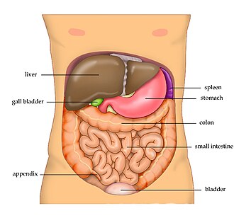 The human male abdomen and organs which can be found beneath the surface | |
| Details | |
| Actions | Movement and support for the torsoAssistance with breathingProtection for the inner organsPostural support |
| Identifiers | |
| Latin | abdomen |
| Greek | ἦτρον |
| MeSH | D000005 |
| TA98 | A01.1.00.016 |
| TA2 | 127 |
| FMA | 9577 |
| Anatomical terminology | |
An abdomen (also belly or stomach[1] in vertebrates, or metasoma in arthropods) is the front part of the torso between the thorax (chest) and pelvis in humans and in other vertebrates. The area occupied by the abdomen is called the abdominal cavity. In arthropods, it is the posteriortagma of the body; it follows the thorax or cephalothorax.[2]
In humans, the abdomen stretches from the thorax at the thoracic diaphragm to the pelvis at the pelvic brim. The pelvic brim stretches from the lumbosacral joint (the intervertebral disc between L5 and S1) to the pubic symphysis and is the edge of the pelvic inlet. The space above this inlet and under the thoracic diaphragm is termed the abdominal cavity. The boundary of the abdominal cavity is the abdominal wall in the front and the peritoneal surface at the rear.
In vertebrates, the abdomen is a large body cavity enclosed by the abdominal muscles, at the front and to the sides, and by part of the vertebral column at the back. Lower ribs can also enclose ventral and lateral walls. The abdominal cavity is continuous with, and above, the pelvic cavity. It is attached to the thoracic cavity by the diaphragm. Structures such as the aorta, inferior vena cava and esophagus pass through the diaphragm. Both the abdominal and pelvic cavities are lined by a serous membrane known as the parietal peritoneum. This membrane is continuous with the visceral peritoneum lining the organs.[3] The abdomen in vertebrates contains a number of organs belonging to, for instance, the digestive system, urinary system, and muscular system.
Contents
ช่องท้องประกอบด้วยอวัยวะส่วนใหญ่ของระบบย่อยอาหารได้แก่กระเพาะอาหารลำไส้เล็กและลำไส้ใหญ่พร้อมไส้ติ่งที่ติดอยู่อวัยวะย่อยอาหารอื่นๆ เรียกว่าอวัยวะเสริมการย่อย อาหาร ได้แก่ตับถุงน้ำดีที่ติดอยู่และตับอ่อนซึ่งเชื่อมต่อกับส่วนอื่นๆ ของระบบผ่านท่อต่างๆ ม้ามและอวัยวะของระบบทางเดินปัสสาวะได้แก่ไตและต่อมหมวกไต ก็อยู่ในช่องท้องเช่นกัน พร้อมกับหลอดเลือดจำนวนมาก ได้แก่ เส้นเลือดแดงใหญ่และเส้นเลือดดำใหญ่ กระเพาะปัสสาวะ มดลูก ท่อนำไข่ และรังไข่ อาจมองได้ว่าเป็นอวัยวะในช่องท้องหรืออวัยวะในอุ้งเชิงกราน สุดท้าย ช่องท้องมีเยื่อหุ้มขนาดใหญ่ที่เรียกว่าเยื่อบุช่องท้องเยื่อบุช่องท้องอาจคลุมอวัยวะบางส่วนอย่างสมบูรณ์ ในขณะที่อาจคลุมเพียงด้านเดียวของอวัยวะที่มักอยู่ใกล้ผนังช่องท้องมากกว่า บริเวณนี้เรียกว่าช่องว่างหลังเยื่อบุช่องท้อง และไตกับท่อปัสสาวะจัดเป็นอวัยวะที่อยู่ในช่องว่างหลังเยื่อบุช่องท้อง
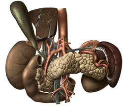 ภาพแสดงอวัยวะและหลอดเลือดต่างๆ ที่อยู่ใกล้กับตับ
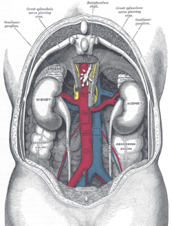 ความสัมพันธ์ระหว่างอวัยวะภายในและหลอดเลือดใหญ่ในช่องท้อง เมื่อมองจากด้านหลัง
กล้ามเนื้อ
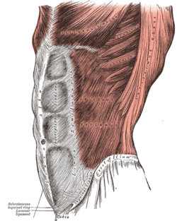

ผนังหน้าท้องมีกล้ามเนื้อสามชั้น เรียงจากด้านนอกเข้าด้านใน ได้แก่กล้ามเนื้อเฉียงด้านนอกกล้ามเนื้อเฉียงด้านในและกล้ามเนื้อหน้าท้องตามขวาง[ 4 ]กล้ามเนื้อสามชั้นแรกทอดยาวระหว่างกระดูกสันหลัง ซี่โครงส่วนล่าง สันกระดูกเชิงกรานและกระดูกหัวหน่าวของสะโพกเส้นใยทั้งหมดรวมกันเข้าหาเส้นกลางลำตัวและล้อมรอบกล้ามเนื้อหน้าท้อง ตรง เป็นปลอกหุ้มก่อนที่จะรวมกันที่ด้านตรงข้ามที่เส้นกลางลำตัวความแข็งแรงได้มาจากการไขว้กันของเส้นใย โดยที่กล้ามเนื้อเฉียงด้านนอกทอดลงและไปข้างหน้ากล้ามเนื้อเฉียงด้านในทอดขึ้นและไปข้างหน้า และกล้ามเนื้อหน้าท้องตามขวางทอดไปข้างหน้าในแนวนอน[ 4 ]
The transverse abdominal muscle is flat and triangular, with its fibers running horizontally. It lies between the internal oblique and the underlying transverse fascia. It originates from the inguinal ligament, costal cartilages 7–12, the iliac crest and thoracolumbar fascia. Inserts into the conjoint tendon, xiphoid process, linea alba and the pubic crest.
The rectus abdominis muscles are long and flat. The muscle is crossed by three fibrous bands called the tendinous intersections. The rectus abdominis is enclosed in a thick sheath, formed as described above, by fibers from each of the three muscles of the lateral abdominal wall. They originate at the pubis bone, run up the abdomen on either side of the linea alba, and insert into the cartilages of the fifth, sixth, and seventh ribs. In the region of the groin, the inguinal canal, is a passage through the layers. This gap is where the testes can drop through the wall and where the fibrous cord from the uterus in the female runs. This is also where weakness can form, and cause inguinal hernias.[4]
The pyramidalis muscle is small and triangular. It is located in the lower abdomen in front of the rectus abdominis. It originates at the pubic bone and is inserted into the linea alba halfway up to the navel.
Function
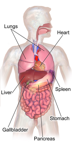
Functionally, the human abdomen is where most of the digestive tract is placed and so most of the absorption and digestion of food occurs here. The alimentary tract in the abdomen consists of the lower esophagus, the stomach, the duodenum, the jejunum, ileum, the cecum and the appendix, the ascending, transverse and descending colons, the sigmoid colon and the rectum. Other vital organs inside the abdomen include the liver, the kidneys, the pancreas and the spleen.
The abdominal wall is split into the posterior (back), lateral (sides), and anterior (front) walls.
Movement, breathing and other functions
กล้ามเนื้อหน้าท้องมีหน้าที่สำคัญหลายอย่าง พวกมันช่วยเป็นกล้ามเนื้อในการหายใจออกในกระบวนการหายใจออกอย่างแรงนอกจากนี้ กล้ามเนื้อเหล่านี้ยังทำหน้าที่ปกป้องอวัยวะภายใน ยิ่งไปกว่านั้น เมื่อรวมกับกล้ามเนื้อหลัง พวกมันยังช่วยพยุงท่าทางและมีความสำคัญในการกำหนดรูปร่าง เมื่อกล่องเสียงปิดและทรวงอกและ กระดูก เชิงกรานคงที่กล้ามเนื้อ เหล่านี้จะมีบทบาทสำคัญในการ ไอ ปัสสาวะอุจจาระการคลอดบุตรอาเจียนและการร้องเพลง[ 4 ] เมื่อกระดูกเชิงกรานคงที่ กล้ามเนื้อ เหล่านี้สามารถเริ่มต้นการเคลื่อนไหวของลำตัวไปข้างหน้าได้ พวกมันยังป้องกันการยืดตัวมากเกินไปเมื่อทรวงอกคงที่ กล้ามเนื้อเหล่านี้สามารถดึงกระดูกเชิงกรานขึ้น และสุดท้าย พวกมันสามารถงอแนวกระดูกสันหลังไปด้านข้างและช่วยในการหมุนของลำตัวได้[ 4 ]
ท่าทาง
กล้ามเนื้อหน้าท้องส่วนลึก (Transverse Abdominis) เป็นกล้ามเนื้อที่อยู่ลึกที่สุด ดังนั้นจึงไม่สามารถสัมผัสได้จากภายนอก กล้ามเนื้อนี้มีผลต่อท่าทางของร่างกายอย่างมาก กล้ามเนื้อหน้าท้องส่วนใน (Internal Obliques) ก็อยู่ลึกเช่นกันและมีผลต่อท่าทางของร่างกายเช่นกัน กล้ามเนื้อทั้งสองส่วนนี้เกี่ยวข้องกับการหมุนและการงอไปด้านข้างของกระดูกสันหลังและใช้ในการงอและพยุงกระดูกสันหลังจากด้านหน้า กล้ามเนื้อหน้าท้องส่วนนอก (External Obliques) อยู่ตื้นกว่าและเกี่ยวข้องกับการหมุนและการงอไปด้านข้างของกระดูกสันหลังเช่นกัน นอกจากนี้ยังช่วยพยุงกระดูกสันหลังเมื่อยืนตรง กล้ามเนื้อหน้าท้องส่วนกลาง (Rectus Abdominis) ไม่ใช่กล้ามเนื้อหน้าท้องที่อยู่ตื้นที่สุด ปลอกเอ็นที่ยื่นออกมาจากกล้ามเนื้อหน้าท้องส่วนนอกจะคลุมกล้ามเนื้อหน้าท้องส่วนกลางไว้ กล้ามเนื้อหน้าท้องส่วนกลางเป็นกล้ามเนื้อที่คนที่มีสุขภาพดีมาก ๆ พัฒนาให้เป็นซิกแพ็ก แม้ว่าจะมีห้าส่วนแนวตั้งในแต่ละด้าน สองส่วนล่างอยู่เหนือกระดูกหัวหน่าวและมักจะมองไม่เห็น หน้าที่ของกล้ามเนื้อหน้าท้องส่วนกลางคือการงอหลังไปข้างหน้า (การงอ) หน้าที่หลักของกล้ามเนื้อหน้าท้องคือการงอกระดูกสันหลังไปข้างหน้าเมื่อหดตัวแบบรวมศูนย์[ 5 ]
สังคมและวัฒนธรรม
ทัศนคติทางสังคมและวัฒนธรรมเกี่ยวกับรูปลักษณ์ภายนอกของหน้าท้องนั้นมีความหมายแตกต่างกันไปทั่วโลก ขึ้นอยู่กับสังคมนั้นๆน้ำหนักเกินอาจถูกมองว่าเป็นตัวบ่งชี้ความมั่งคั่งและเกียรติยศเนื่องจากการรับประทานอาหารมากเกินไป หรือเป็นสัญญาณของสุขภาพที่ไม่ดีเนื่องจากการขาดการออกกำลังกาย ในหลายวัฒนธรรม หน้าท้องที่เปลือยเปล่าถูกมองว่าเป็นเรื่องทางเพศอย่างชัดเจนและถูกมองในลักษณะเดียวกับ การโชว์ เนินอก
ออกกำลังกาย

เนื่องจากกล้ามเนื้อหน้าท้องเป็นองค์ประกอบสำคัญในการรองรับกระดูกสันหลังและมีส่วนช่วยให้มีท่าทางที่ดี จึงจำเป็นอย่างยิ่งที่จะต้องออกกำลังกายกล้ามเนื้อหน้าท้องควบคู่ไปกับกล้ามเนื้อหลังอย่างเหมาะสม เพราะเมื่อกล้ามเนื้อเหล่านี้อ่อนแอหรือตึงเกินไป อาจทำให้เกิดอาการปวดเกร็งและบาดเจ็บได้เมื่อออกกำลังกายอย่างเหมาะสม กล้ามเนื้อหน้าท้องจะช่วยปรับปรุงท่าทางและการทรงตัว ลดโอกาส การเกิดอาการ ปวดหลังลดความรุนแรงของอาการปวดหลัง[ 7 ]ป้องกันการบาดเจ็บ[ 8 ]ช่วยหลีกเลี่ยงการผ่าตัดหลังบางประเภท และช่วยในการรักษาปัญหาเกี่ยวกับหลังหรือหลังการผ่าตัดกระดูกสันหลัง เมื่อกล้ามเนื้อหน้าท้องแข็งแรงขึ้น ก็จะช่วยเพิ่มความยืดหยุ่นได้เช่นกัน กล้ามเนื้อหน้าท้องสามารถบริหารได้ด้วยการออกกำลังกายเพื่อเพิ่มความแข็งแรงและความฟิต และผ่านการฝึกฝนวินัยต่างๆ ที่ช่วยเพิ่มความแข็งแรงของร่างกายโดยทั่วไป เช่นพิลาทิส [ 9 ] โยคะ [ 10 ] ไทชิและการวิ่งจ็อกกิ้ง
ความสำคัญทางคลินิก
ภาวะอ้วนลงพุงคือภาวะที่ไขมันหน้าท้องหรือไขมันในช่องท้องสะสมมากเกินไประหว่างอวัยวะภายในช่องท้อง ซึ่งเกี่ยวข้องกับความเสี่ยงที่สูงขึ้นของโรคหัวใจโรคหอบหืดและโรคเบาหวานชนิดที่ 2
การบาดเจ็บที่ช่องท้องเป็นการบาดเจ็บที่ช่องท้องและอาจเกี่ยวข้องกับความเสียหายต่ออวัยวะในช่องท้อง มีความเสี่ยงที่จะเกิดการเสียเลือดมากและการติดเชื้อ[ 11 ]การบาดเจ็บที่หน้าอกส่วนล่างอาจทำให้เกิดการบาดเจ็บที่ม้ามและตับ[ 12 ]
ภาวะท้องยุบเข้าด้านในเรียกว่าท้องสแคฟอยด์[ 13 ]ในทารกแรกเกิด อาจบ่งชี้ถึงไส้เลื่อนกระบังลม [ 14 ] โดยทั่วไปแล้ว มักบ่งชี้ถึงภาวะทุพโภชนาการ[ 15 ]
โรค
โรคระบบทางเดินอาหารหลายชนิดส่งผลกระทบต่ออวัยวะในช่องท้อง ซึ่งรวมถึงโรคกระเพาะอาหารโรคตับโรคตับอ่อน โรคถุงน้ำดีและท่อน้ำดีส่วนโรคที่เกี่ยวกับลำไส้ ได้แก่ โรคลำไส้อักเสบโรคเซลิแอค โรคถุงผนังลำไส้อักเสบและกลุ่มอาการลำไส้แปรปรวน
การตรวจสอบ
สามารถใช้ ขั้นตอนทางการแพทย์ต่างๆในการตรวจอวัยวะในระบบทางเดินอาหารได้ ซึ่งได้แก่การส่องกล้องตรวจลำไส้เล็กและลำไส้ใหญ่ การส่องกล้องตรวจลำไส้ใหญ่ส่วนปลาย การส่องกล้องตรวจลำไส้เล็ก การส่องกล้องตรวจหลอดอาหาร กระเพาะอาหาร และลำไส้เล็กส่วนต้นและการส่องกล้องตรวจลำไส้ใหญ่เสมือนจริง นอกจากนี้ ยังมี เทคนิค การถ่ายภาพทางการแพทย์ อีกหลายอย่าง ที่สามารถนำมาใช้ได้ จุดสังเกตบนพื้นผิวมีความสำคัญในการตรวจช่องท้อง
จุดสังเกตบนพื้นผิว

บริเวณกึ่งกลางลำตัว จะมีร่องเล็กๆ ทอดยาวจาก กระดูกอก ส่วนบน(xiphoid process) ไปจนถึงกระดูกหัวหน่าว ส่วนล่าง ( pubic symphysis) ซึ่งแสดงถึง เส้นกลางลำตัว ( linea alba ) บริเวณกึ่งกลางของเส้นกลางลำตัวนี้คือ สะดือกล้าม เนื้อหน้า ท้อง (rectus abdominis)ที่อยู่ด้านข้างของเส้นกลางลำตัวจะเห็นได้ชัดเจนในคนที่มีกล้ามเนื้อมาก ขอบของกล้ามเนื้อเหล่านี้ถูกขัดจังหวะด้วยรอยบุ๋มตามขวางสามรอยขึ้นไป ซึ่งแสดงถึงจุดตัดของเอ็นกล้ามเนื้อโดยปกติจะมีหนึ่งรอยอยู่เหนือกระดูกอก หนึ่งรอยอยู่ที่สะดือ และอีกหนึ่งรอยอยู่ตรงกลาง การรวมกันของเส้นกลางลำตัวและจุดตัดของเอ็นกล้ามเนื้อนี้เองที่ทำให้เกิด "ซิกแพ็ก" หน้าท้องที่หลายคนปรารถนา
ขอบบนด้านข้างของช่องท้องคือขอบใต้ซี่โครง (ที่หรือใกล้ระนาบใต้ซี่โครง ) ซึ่งเกิดจากกระดูกอ่อนของซี่โครงเทียม (8, 9, 10) ที่เชื่อมต่อกัน ขอบล่างด้านข้างคือสันด้านหน้าของกระดูกเชิงกรานและเอ็นของปูพาร์ตซึ่งทอดจากกระดูกสันหลังส่วนหน้าด้านบนของกระดูกเชิงกรานไปยังกระดูกสันหลังของกระดูกหัวหน่าวขอบล่างเหล่านี้มีร่องที่มองเห็นได้ชัดเจน เหนือกระดูกสันหลังของกระดูกหัวหน่าวทั้งสองข้างคือวงแหวนช่องท้องภายนอก ซึ่งเป็นช่องเปิดในผนังกล้ามเนื้อของช่องท้องที่ท่ออสุจิโผลออกมาในเพศชาย และเป็นช่องที่ไส้เลื่อนขาหนีบอาจแตกได้
วิธีหนึ่งที่จะช่วยระบุตำแหน่งของอวัยวะภายในช่องท้องได้คือ การลากเส้นแนวนอนสามเส้นและเส้นแนวตั้งสองเส้น
เส้นแนวนอน
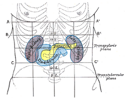
- The highest of the former is the transpyloric line of C. Addison, which is situated halfway between the suprasternal notch and the top of the pubic symphysis, and often cuts the pyloric opening of the stomach an inch to the right of the mid-line. The hilum of each kidney is a little below it, while its left end approximately touches the lower limit of the spleen. It corresponds to the first lumbar vertebra behind.
- The second line is the subcostal line, drawn from the lowest point of the subcostal arch (tenth rib). It corresponds to the upper part of the third lumbar vertebra, and it is an inch or so above the umbilicus. It indicates roughly the transverse colon, the lower ends of the kidneys, and the upper limit of the transverse (3rd) part of the duodenum.
- The third line is called the intertubercular line, and runs across between the two rough tubercles, which can be felt on the outer lip of the crest of the ilium about two and a half inches (64 mm) from the anterior superior spine. This line corresponds to the body of the fifth lumbar vertebra, and passes through or just above the ileo-caecal valve, where the small intestine joins the large intestine.
Vertical lines
The two vertical or mid-Poupart lines are drawn from the point midway between the anterior superior spine and the pubic symphysis on each side, vertically upward to the costal margin.
- The right one is the most valuable, as the ileo-caecal valve is situated where it cuts the intertubercular line. The orifice of the appendix lies an inch lower, at McBurney's point. In its upper part, the vertical line meets the transpyloric line at the lower margin of the ribs, usually the ninth, and here the gallbladder is situated.
- The left mid-Poupart line corresponds in its upper three-quarters to the inner edge of the descending colon.
The right subcostal margin corresponds to the lower limit of the liver, while the right nipple is about half an inch above its upper limit.
Quadrants and regions
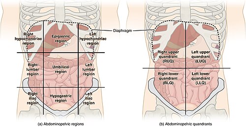
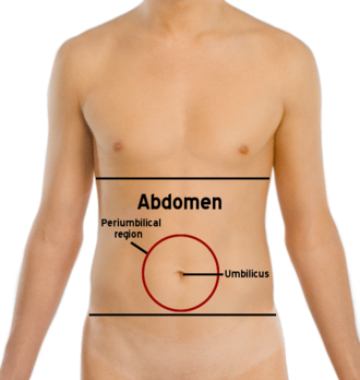
The abdomen can be divided into quadrants or regions to describe the location of an organ or structure. Classically, quadrants are described as the left upper, left lower, right upper, and right lower. Quadrants are also often used in describing the site of an abdominal pain.[16]
The abdomen can also be divided into nine regions.
| right hypochondriac/hypochondrium | epigastric/epigastrium | left hypochondriac/hypochondrium |
| right lumbar/flank/latus/lateral | umbilical | left lumbar/flank/latus/lateral |
| right inguinal/iliac | hypogastric/suprapubic | left inguinal/iliac |
These terms stem from "hypo" meaning "below" and "epi" means "above", while "chondron" means "cartilage" (in this case, the cartilage of the rib) and "gaster" means stomach. The reversal of "left" and "right" is intentional, because the anatomical designations reflect the patient's own right and left.)
The "right iliac fossa" (RIF) is a common site of pain and tenderness in patients who have appendicitis. The fossa is named for the underlying iliac fossa of the hip bone, and thus is somewhat imprecise. Most of the anatomical structures that will produce pain and tenderness in this region are not in fact in the concavity of the ileum. However, the term is in common usage.
Across animal phyla and classes
Chordata
Mammals
Abdominal organs can be highly specialized in some mammals. For example, the stomach of ruminants, (a suborder of mammals that includes cattle and sheep), is divided into four chambers – rumen, reticulum, omasum and abomasum.[17]
Arthropoda
In arthropods, the abdomen is built up of a series of upper plates known as tergites and lower plates known as sternites, the whole being held together by a tough yet stretchable membrane.
Insects
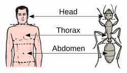

In insects, the abdomen contains the insect's digestive tract and reproductive organs. It consists of eleven segments in most orders of insects, though the eleventh segment is absent in the adult of most higher orders. The number of these segments does vary from species to species with the number of segments visible reduced to only seven in the common honey bee. In the Collembola (springtails), the abdomen has only six segments.
บางครั้งช่องท้องก็มีการเปลี่ยนแปลงอย่างมาก ในApocrita (ผึ้ง มด และแตน) ปล้องแรกของช่องท้องจะเชื่อมติดกับทรวงอกและเรียกว่าpropodeumในมดปล้องที่สองจะก่อตัวเป็นpetiole ที่แคบ มดบางชนิดมีปล้อง postpetioleเพิ่มเติมและปล้องที่เหลือจะก่อตัวเป็นgasterที่ มีลักษณะเป็นกระเปาะ [ 18 ] petiole และ gaster (ปล้องช่องท้องที่ 2 เป็นต้นไป) เรียกรวมกันว่าmetasoma
แตกต่างจากสัตว์ขาปล้องชนิดอื่นๆ แมลงไม่มีขาที่ส่วนท้องในระยะโตเต็มวัย แม้ว่ากลุ่มProturaจะมีระยางค์คล้ายขาที่เจริญไม่เต็มที่บนปล้องท้องสามปล้องแรก และกลุ่ม Archaeognathaมี "สไตลัส" ขนาดเล็กที่มีข้อต่อ ซึ่งบางครั้งก็ถือว่าเป็นระยางค์ที่เจริญไม่เต็มที่เช่นกัน ตัวอ่อนของแมลงหลายชนิด รวมถึงกลุ่มLepidopteraและSymphyta (แมลงเลื่อย) มีระยางค์เนื้อนุ่มที่เรียกว่าขาเทียมบนปล้องท้อง (รวมถึงขาอกที่คุ้นเคยมากกว่า) ซึ่งช่วยให้พวกมันเกาะขอบใบพืชขณะเดินไปมาได้
แมงมุม
ในสัตว์กลุ่มแมงมุม (แมงมุม แมงป่อง และญาติๆ) คำว่า "ท้อง" ใช้แทนกันได้กับคำว่า " โอพิสโทโซมา " ("ส่วนท้ายลำตัว") ซึ่งเป็นส่วนของร่างกายที่อยู่ด้านหลังส่วนที่มีขาและหัว (โพรโซมาหรือเซฟาโลทอแรกซ์ )
ดูเพิ่มเติม
ลิงก์ภายนอก

- .สารานุกรมเล่มใหม่ของคอลลิเออร์ . 1921.
 สื่อที่เกี่ยวข้องกับช่องท้องในวิกิมีเดียคอมมอนส์